
Page 24 - Read Online
P. 24
Page 8 of 11 Tran et al. J Cancer Metastasis Treat 2020;6:47 I http://dx.doi.org/10.20517/2394-4722.2020.104
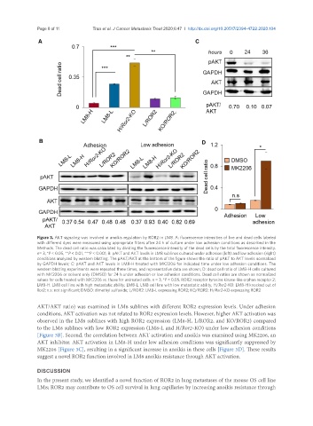
Figure 3. AKT signaling was involved in anoikis regulation by ROR2 in LM8. A: fluorescence intensities of live and dead cells labeled
with different dyes were measured using appropriate filters after 24 h of culture under low adhesion conditions as described in the
Methods. The dead cell ratio was calculated by dividing the fluorescence intensity of the dead cells by the total fluorescence intensity.
n= 3, *P < 0.05, **P < 0.01, ***P < 0.001; B: pAKT and AKT levels in LM8 sublines cultured under adhesion (left) and low adhesion (right)
conditions analyzed by western blotting. The pAKT/AKT at the bottom of the figure shows the ratio of pAKT to AKT levels normalized
by GAPDH levels; C: pAKT and AKT levels in LM8-H treated with MK2206 for indicated time under low adhesion conditions. The
western blotting experiments were repeated three times, and representative data are shown; D: dead cell ratio of LM8-H cells cultured
with MK2206 or solvent only (DMSO) for 24 h under adhesion or low adhesion conditions. Dead cell ratios are shown as normalized
values for cells treated with MK2206 vs. those for untreated cells. n = 3, *P < 0.05. ROR2: receptor tyrosine kinase-like orphan receptor 2;
LM8-H: LM8 cell line with high metastatic ability; LM8-L: LM8 cell line with low metastatic ability; H/Ror2-KO: LM8-H knocked out of
Ror2; n.s: not significant; DMSO: dimethyl sulfoxide; L/ROR2: LM8-L expressing ROR2; KO/ROR2: H/Ror2-KO expressing ROR2
AKT/AKT ratio) was examined in LM8 sublines with different ROR2 expression levels. Under adhesion
conditions, AKT activation was not related to ROR2 expression levels. However, higher AKT activation was
observed in the LM8 sublines with high ROR2 expression (LM8-H, L/ROR2, and KO/ROR2) compared
to the LM8 sublines with low ROR2 expression (LM8-L and H/Ror2-KO) under low adhesion conditions
[Figure 3B]. Second, the correlation between AKT activation and anoikis was examined using MK2206, an
AKT inhibitor. AKT activation in LM8-H under low adhesion conditions was significantly suppressed by
MK2206 [Figure 3C], resulting in a significant increase in anoikis in these cells [Figure 3D]. These results
suggest a novel ROR2 function involved in LM8 anoikis resistance through AKT activation.
DISCUSSION
In the present study, we identified a novel function of ROR2 in lung metastases of the mouse OS cell line
LM8; ROR2 may contribute to OS cell survival in lung capillaries by increasing anoikis resistance through