
Page 22 - Read Online
P. 22
Su et al. J Cancer Metastasis Treat 2020;6:19 I http://dx.doi.org/10.20517/2394-4722.2020.48 Page 17 of 21
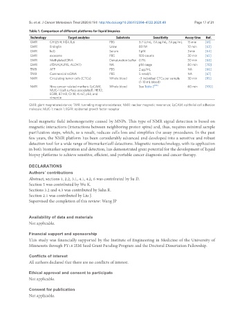
Table 1. Comparison of different platforms for liquid biopsies
Technology Target analytes Substrate Sensitivity Assay time Ref.
GMR CA125 II, HE4, IL6 PBS 3.7 U/mL, 7.4 pg/mL, 7.4 pg/mL 15 min [61]
GMR Endoglin Urine 83 fM 10 min [63]
GMR hcG Serum 1 pM 3 min [64]
GMR exosome PBS 100 counts 30 min [65]
GMR Methylated DNA Denaturation buffer 0.1% 30 min [68]
GMR cfDNA(AU115, AU247) NA pM range 80 min [70]
TMR AFP PBS 2 μg/mL NA [66]
TMR Commercial ssDNA PBS 5 nmol/L NA [67]
NMR Circulating tumor cells (CTCs) Whole blood ~3 individual CTCs per sample 30 min [95]
(1-10 mL blood)
NMR Nine cancer-related markers: EpCAM, Whole blood See Table 2 [95] 60 min [100]
MUC-1 (cell surface associated), HER2,
EGFR, B7-H3, CK18, Ki-67, p53, and
vimentin
GMR: giant magnetoresistance; TMR: tunneling magnetoresistance; NMR: nuclear magnetic resonance; EpCAM: epithelial cell adhesion
molecule; MUC-1: mucin 1; EGFR: epidermal growth factor receptor
local magnetic field inhomogeneity caused by MNPs. This type of NMR signal detection is based on
magnetic interactions (interactions between neighboring proton spins) and, thus, requires minimal sample
purification steps, which, as a result, reduces cells loss and simplifies the assay procedures. In the past
few years, the NMR platform has been considerably advanced and developed into a sensitive and robust
detection tool for a wide range of biomarker/cell detections. Magnetic nanotechnology, with its application
in both biomarker separation and detection, has demonstrated great potential for the development of liquid
biopsy platforms to achieve sensitive, efficient, and portable cancer diagnosis and cancer therapy.
DECLARATIONS
Authors’ contributions
Abstract, sections 1, 2.2, 3.1, 4.1, 4.2, 6 was contributed by Su D.
Section 5 was contributed by Wu K.
Sections 3.2 and 4.3 was contributed by Saha R.
Section 2.1 was contributed by Liu J.
Supervised the completion of this review: Wang JP
Availability of data and materials
Not applicable.
Financial support and sponsorship
This study was financially supported by the Institute of Engineering in Medicine of the University of
Minnesota through FY18 IEM Seed Grant Funding Program and the Doctoral Dissertation Fellowship.
Conflicts of interest
All authors declared that there are no conflicts of interest.
Ethical approval and consent to participate
Not applicable.
Consent for publication
Not applicable.