
Page 112 - Read Online
P. 112
Vidoni et al. J Cancer Metastasis Treat 2021;7:4 I http://dx.doi.org/10.20517/2394-4722.2020.95 Page 5 of 20
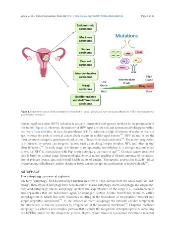
Figure 3. Endometrial cancer. Brief presentation of endometrial cancer groups and main molecular alterations. HER2: human epidermal
growth factor receptor 2
human papilloma virus (HPV) infection is sexually transmitted and appears involved in the progression of
this tumor [Figure 4]. However, the majority of HPV types are low-risk and spontaneously disappear within
two years from infection. In fact, the prevalence of HPV-infection is high in women of under 25 years of
[35]
age, whereas the peak of cervical cancer death occurs in middle-aged women . HPV 16 and 18 are the
[36]
most common oncogenic genotypes found in 75% of invasive cervical carcinoma . The tumor progression
is influenced by several carcinogenic factors, such as smoking, herpes simplex, HIV, and other genital
[37]
virus infections . In early stage, this disease is asymptomatic; nevertheless, it is strongly recommended
[38]
to test for HPV in conjunction with Pap smear cytology at 21 years of age . Cervical cancer treatment
plan is based on clinical stage, histopathological type of tumor, grading of disease, presence of metastasis,
size of primary lesion, age, and overall health status of patient. Therapeutic approaches include radical
hysterectomy, radiotherapy, and/or platinum-based chemotherapy, in combination or independently .
[34]
AUTOPHAGY
The autophagy process at a glance
The term “autophagy” first described by Christian De Duve in 1963, derives from the Greek word for “self-
eating”. Three types of autophagy have been described: macro-autophagy, micro-autophagy, and chaperone-
mediated autophagy. Macro-autophagy involves the sequestration of the cargo (i.e., macromolecules
and organelles that are redundant, aged, or damaged) within double membrane vacuoles, called
autophagosomes, which fuse with lysosomes resulting in the breakdown of encapsulated material into
simple recyclable components . In the instance of micro-autophagy, the cytosolic cellular components
[39]
[40]
are internalized within the lysosomes by invagination of the lysosomal membrane . Chaperon-mediated
autophagy is a selective and complex pathway that includes the recognition of targeted protein, containing
the KFERQ motif, by the chaperone protein Hsp70, which binds to lysosomal membrane receptor