
Page 102 - Read Online
P. 102
Page 6 of 11 Tessari et al. J Cancer Metastasis Treat 2020;6:18 I http://dx.doi.org/10.20517/2394-4722.2020.32
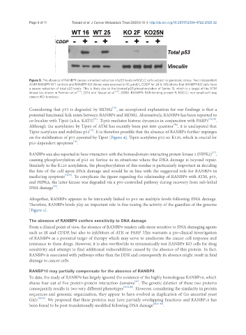
Figure 3. The absence of RANBP9 causes a marked reduction of p53 levels in NSCLC cells subject to genotoxic stress. Two independent
A549 RANBP9 WT controls and RANBP9 KO clones were exposed to 10 μmol/L CDDP for 24 h. WB shows that RANBP9 KO cells have
a severe reduction of total p53 levels. This is likely due to the blunted p53 phosphorylation of Serine 15, which is a target of the ATM
kinase (as shown in Palmieri et al. [56] , 2016 and Tessari et al. [50] , 2018). RANBP9: RAN binding protein 9; NSCLC: non-small cell lung
cancer; KO: knockout
[70]
Considering that p53 is degraded by MDM2 , an unexplored explanation for our findings is that a
potential functional link exists between RANBP9 and MDM2. Alternatively, RANBP9 has been reported to
[71]
co-localize with Tip60 (a.k.a. KAT5) . Tip60 mediates histone dynamics in conjunction with PARP1 [72,73] .
[74]
Although the acetylation by Tip60 of ATM has recently been put into question , it is undisputed that
[75]
Tip60 acetylates and stabilizes p53 . It is therefore possible that the absence of RANBP9 further impinges
on the stabilization of p53 operated by Tip60 [Figure 4]. Tip60 acetylates p53 on K120, which is crucial for
p53-dependent apoptosis .
[76]
[77]
RANBP9 was also reported to have interaction with the homeodomain-interacting protein kinase 2 (HIPK2) ,
causing phosphorylation of p53 on Serine 46 in situations where the DNA damage is beyond repair.
Similarly to the K120 acetylation, the phosphorylation of this residue is particularly important in deciding
the fate of the cell upon DNA damage and would be in line with the suggested role for RANBP9 in
mediating apoptosis [40,78] . To complicate the jigsaw regarding the relationship of RANBP9 with ATM, p53,
and HIPK2, the latter kinase was degraded via a p53-controlled pathway during recovery from sub-lethal
[79]
DNA damage .
Altogether, RANBP9 appears to be intricately linked to p53 on multiple levels following DNA damage.
Therefore, RANBP9 levels play an important role in fine-tuning the activity of the guardian of the genome
[Figure 4].
The absence of RANBP9 confers sensitivity to DNA damage
From a clinical point of view, the absence of RANBP9 renders cells more sensitive to DNA damaging agents
such as IR and CDDP, but also to inhibitors of ATR or PARP. This warrants a pre-clinical investigation
of RANBP9 as a potential target of therapy which may serve to ameliorate the cancer cell response and
resistance to these drugs. However, it is also worthwhile to systematically test RANBP9 KO cells for drug
sensitivity and attempt to find additional vulnerabilities caused by the absence of this protein. In fact,
RANBP9 is associated with pathways other than the DDR and consequently its absence might result in fatal
damage to cancer cells.
RANBP10 may partially compensate for the absence of RANBP9
To date, the study of RANBP9 has largely ignored the existence of the highly homologous RANBP10, which
[47]
shares four out of five protein-protein interaction domains . The genetic deletion of these two proteins
consequently results in two very different phenotypes [35,36,80] . However, considering the similarity in protein
sequences and genomic organization, they appear to have evolved as duplication of the ancestral yeast
Gid1 [28,29] . We proposed that these proteins may have partially overlapping functions and RANBP10 has
been found to be post-translationally modified following DNA damage [66,81-83] .