
Page 280 - Read Online
P. 280
Mohamedi et al. J Cancer Metastasis Treat 2019;5:37 I http://dx.doi.org/10.20517/2394-4722.2018.81 Page 11 of 15
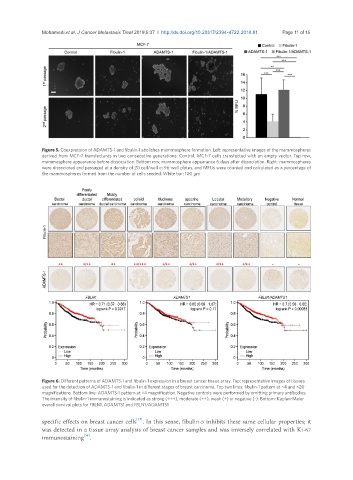
Figure 5. Coexpression of ADAMTS-1 and fibulin-1 abolishes mammosphere formation. Left: representative images of the mammospheres
derived from MCF-7 transfectants in two consecutive generations. Control, MCF-7 cells transfected with an empty vector. Top row,
mammosphere appearance before dissociation. Bottom row, mammosphere appearance 6 days after dissociation. Right: mammospheres
were dissociated and passaged at a density of 20 cell/well in 96-well plates, and MFUs were counted and calculated as a percentage of
the mammospheres formed from the number of cells seeded. White bar: 100 μm
Figure 6. Different patterns of ADAMTS-1 and fibulin-1 expression in a breast cancer tissue array. Top: representative images of tissues
used for the detection of ADAMTS-1 and fibulin-1 in different stages of breast carcinoma. Top two lines: fibulin-1 pattern at ×4 and ×20
magnifications. Bottom line: ADAMTS-1 pattern at ×4 magnification. Negative controls were performed by omitting primary antibodies.
The intensity of fibulin-1 immunostaining is indicated as strong (+++), moderate (++), weak (+) or negative (-). Bottom: Kaplan-Meier
overall survival plots for FBLN1, ADAMTS1 and FBLN1/ADAMTS1
[37]
specific effects on breast cancer cells . In this sense, fibulin-5 inhibits these same cellular properties; it
was detected in a tissue array analysis of breast cancer samples and was inversely correlated with Ki-67
[38]
immunostaining .