
Page 279 - Read Online
P. 279
Page 10 of 15 Mohamedi et al. J Cancer Metastasis Treat 2019;5:37 I http://dx.doi.org/10.20517/2394-4722.2018.81
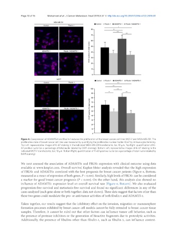
Figure 4. Coexpression of ADAMTS-1 and fibulin-1 reduces the proliferation of the breast cancer cell lines MCF-7 and MDA-MB-231. The
proliferative state of breast cancer cell lines was measured by quantifying the proliferative nuclear marker Ki-67 by immunocytochemistry.
Top Left: representative images of Ki-67 staining in the indicated MDA-MB-231 transfectants; bar, 10 μm. Top Right: quantification of Ki-
67-positive nuclei (as a percentage of total nuclei labeled by DAPI staining). Bottom Left, representative images of Ki-67 staining in the
indicated MCF-7 transfectants; bar, 10 μm. Bottom Right, quantification of Ki-67-positive nuclei (as a percentage of total nuclei labeled by
DAPI staining)
We next assessed the association of ADAMTS1 and FBLN1 expression with clinical outcome using data
available at www.kmplot.com. Overall survival Kaplan-Meier analysis revealed that the high expression
of FBLN1 and ADAMTS1 correlated with the best prognosis for breast cancer patients (Figure 6, Bottom;
measured as a mean of expression of both genes, P < 0.001). Similarly, high levels of FBLN1 can be considered
a marker for good breast cancer prognosis (P < 0.005). On the other hand, this analysis also showed no
influence of ADAMTS1 expression level on overall survival rate [Figure 6, Bottom]. We also evaluated
progression-free survival and metastasis-free survival and found no significant differences in any of the
cases analyzed (each gene alone or both together; data not shown). These data suggest that factors other than
these two genes could modulate the pro- or anti-tumor activities of both fibulin-1 and ADAMTS-1.
Taken together, our results suggest that the inhibitory effect on the invasion, migration or mammosphere
formation processes exhibited by breast cancer cell models cannot be fully extended to breast cancer tissue
samples. Therefore, it cannot be ruled out that other factors can influence tumor cell behavior, such as
the presence of protease inhibitors or the generation of bioactive fragments due to proteolytic activities.
Additionally, the presence of fibulins other than fibulin-1, such as fibulin-5, can influence context-