
Page 276 - Read Online
P. 276
Mohamedi et al. J Cancer Metastasis Treat 2019;5:37 I http://dx.doi.org/10.20517/2394-4722.2018.81 Page 7 of 15
A
B
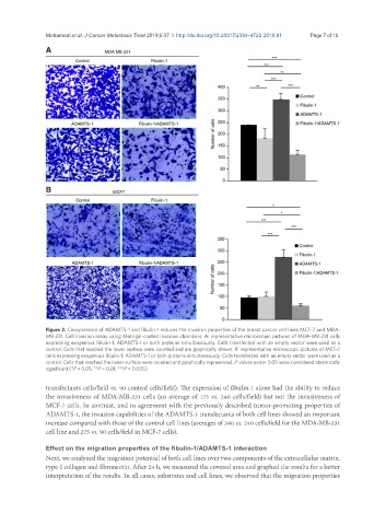
Figure 2. Coexpression of ADAMTS-1 and fibulin-1 reduces the invasion properties of the breast cancer cell lines MCF-7 and MDA-
MB-231. Cell invasion assay using Matrigel-coated invasion chambers. A: representative microscopic pictures of MDA-MB-231 cells
expressing exogenous fibulin-1, ADAMTS-1 or both proteins simultaneously. Cells transfected with an empty vector were used as a
control. Cells that reached the lower surface were counted and are graphically shown; B: representative microscopic pictures of MCF-7
cells expressing exogenous fibulin-1, ADAMTS-1 or both proteins simultaneously. Cells transfected with an empty vector were used as a
control. Cells that reached the lower surface were counted and graphically represented. P values under 0.05 were considered statistically
significant (*P < 0.05, **P < 0.01, ***P < 0.005)
transfectants cells/field vs. 90 control cells/field). The expression of fibulin-1 alone had the ability to reduce
the invasiveness of MDA-MB-231 cells (an average of 175 vs. 240 cells/field) but not the invasiveness of
MCF-7 cells. In contrast, and in agreement with the previously described tumor-promoting properties of
ADAMTS-1, the invasion capabilities of the ADAMTS-1 transfectants of both cell lines showed an important
increase compared with those of the control cell lines (averages of 340 vs. 240 cells/field for the MDA-MB-231
cell line and 275 vs. 90 cells/field in MCF-7 cells).
Effect on the migration properties of the fibulin-1/ADAMTS-1 interaction
Next, we analyzed the migration potential of both cell lines over two components of the extracellular matrix,
type-I collagen and fibronectin. After 24 h, we measured the covered area and graphed the results for a better
interpretation of the results. In all cases, substrates and cell lines, we observed that the migration properties