
Page 158 - Read Online
P. 158
Page 6 of 17 Mooney et al. J Cancer Metastasis Treat 2019;5:19 I http://dx.doi.org/10.20517/2394-4722.2018.93
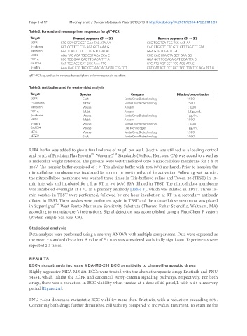
Table 2. Forward and reverse primer sequences for qRT-PCR
Target Forward sequence (5’ → 3’) Reverse sequence (5’ → 3’)
EGFR CTC CCA GTG CCT GAA TAC ATA AA CCG TGG TCA TGC TCC AAT AA
β-catenin GCT CCT TCT CTG AGT GGT AAA G CAC CTG GTC CTC GTC ATT TAG CTT GTA
Vimentin GAT TCA CTC CCT CTG GTT GAT AC GGA GTG TCG GTT GTT
NKD2 AGA TAC ACA TGC CGT ACA CCA C CGG CAG GTA GTA GCT GAA GG
TGF-α CCC TGC GAA GAC TTG AGA TTT A GGA GCT TGC AGA GAT GGA TTA G
GAPDH GAT TCC ACC CAT GGC AAA TTC GTC ATG AGT CCT TCC ACG ATA C
β-actin AAA GAC CTG TAC GCC AAC ACA GTG CTG TCT CGT CAT ACT CCT GCT TGC TGA TCC ACA TCT G
qRT-PCR: quantitative reverse transcription polymerase chain reaction
Table 3. Antibodies used for western blot analysis
Target Species Company Dilution/concentration
EGFR Goat Santa Cruz Biotechnology 1:500
E-cadherin Rabbit Santa Cruz Biotechnology 1:500
Vimentin Mouse Abcam 1:1000
TGF-α Rabbit Abcam 0.2 μg/mL
β-catenin Mouse Santa Cruz Biotechnology 1 μg/mL
NKD2 Rabbit Abcam 1:500
β-actin Mouse Santa Cruz Biotechnology 1:1000
GAPDH Mouse Life Technologies 1 μg/mL
pERK Mouse Santa Cruz Biotechnology 1:500
pEGFR Goat Santa Cruz Biotechnology 1:500
RIPA buffer was added to give a final volume of 20 µL per well. β-actin was utilized as a loading control
TM
TM
and 10 µL of Precision Plus Protein WesternC Standards (BioRad, Hercules, CA) was added to a well as
a molecular weight reference. The proteins were wet-transferred onto a nitrocellulose membrane for 1 h at
100V. The transfer buffer consisted of 1 × Tris-glycine buffer with 20% (v/v) methanol. Prior to transfer, the
nitrocellulose membrane was incubated for 10 min in 100% methanol for activation. Following wet transfer,
the nitrocellulose membrane was washed three times in Tris-buffered saline and Tween 20 (TBST) in 15-
min intervals and incubated for 1 h at RT in 5% (w/v) BSA diluted in TBST. The nitrocellulose membrane
was incubated overnight at 4 ºC in a primary antibody [Table 3], which was diluted in TBST. Three 15-
min washes in TBST were performed, followed by one-hour incubation at RT in a secondary antibody
diluted in TBST. Three washes were performed again in TBST and the nitrocellulose membrane was placed
TM
in Supersignal West Femto Maximum Sensitivity Substrate (Thermo Fisher Scientific, Waltham, MA)
according to manufacturer’s instructions. Signal detection was accomplished using a FluorChem E system
(Protein Simple, San Jose, CA).
Statistical analysis
Data analyses were performed using a one-way ANOVA with multiple comparisons. Data were expressed as
the mean ± standard deviation. A value of P < 0.05 was considered statistically significant. Experiments were
repeated 2-3 times.
RESULTS
ESC-microstrands increase MDA-MB-231 BCC sensitivity to chemotherapeutic drugs
Highly aggressive MDA-MB-231 BCCs were treated with the chemotherapeutic drugs Erlotinib and PNU
74654, which inhibit the EGFR and canonical Wnt/β-catenin signaling pathways, respectively. For both
drugs, there was a reduction in BCC viability when treated at a dose of 20 µmol/L with a 24-h recovery
period [Figure 2A].
PNU 74654 decreased metastatic BCC viability more than Erlotinib, with a reduction exceeding 30%.
Combining both drugs further diminished cell viability compared to individual treatment. To examine the