
Page 162 - Read Online
P. 162
Page 10 of 17 Mooney et al. J Cancer Metastasis Treat 2019;5:19 I http://dx.doi.org/10.20517/2394-4722.2018.93
A B
C D
A
E F
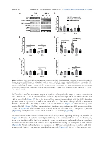
Figure 5. Western blot analysis of long-term protein expression of key EGFR signaling pathway molecules. Metastatic MDA-MB-231
breast cancer cells (BCCs) were co-cultured with embryonic stem cell (ESC)-microstrands for 48 h, followed by removing ESC-
microstrands, replacing BCC media, and analyzing protein expression at 72 h and 120 h. Western blots for protein expression in
metastatic MDA-MB-231 BCCs (CC: co-cultured with ESC-microstrands; E: co-cultured with empty microstrands; C: non-co-cultured
control) (A); densitometry of expression of EGFR (B), precursor TGF-α (C), mature TGF-α (D), pEGFR (E), and pERK (F) (***P < 0.001;
**P < 0.01; *P < 0.05)
BCC media to see if there are other long-term signaling pathway-related changes in protein expression in
MDA-MB-231 BCCs. The BCCs recovered for either one day or three days, which are denoted as 72 h and
120 h, respectively. Figure 5A shows the immunoblots for proteins associated with the EGFR signaling
pathway. Contrasting to results for 48 h of co-culture, after 72 h, there was no change in EGFR expression in
the MDA-MB-231 BCCs following co-culture with ESC-microstrands [Figure 5B]. Precursor TGF-α levels
declined by 72 h [Figure 5C]. There was a statistically significant increase in mature TGF-α expression at the
72-h mark [Figure 5D], which was enhanced by 120 h. There was a decrease after 72 h in pEGFR expression
[Figure 5E]. Finally, there was reduced pERK by 72-h and 120-h marks [Figure 5F].
Immunoblots for molecules related to the canonical Wnt/β-catenin signaling pathway are provided in
Figure 6A. Precursor E-cadherin was not present in any of the samples until 120 h, and for that reason,
there is no band in the blot for the 72-h point. β-catenin expression was unchanged following co-culture
with ESC-microstrands after 72 h, however, it was significantly reduced by 120 h compared to both controls
[Figure 6B]. At 72 h and 120 h, there was an upsurge in NKD2 expression following co-culture with ESC-
microstrands that was significant compared to both controls [Figure 6C]. By 120 h, there was precursor