
Page 157 - Read Online
P. 157
Mooney et al. J Cancer Metastasis Treat 2019;5:19 I http://dx.doi.org/10.20517/2394-4722.2018.93 Page 5 of 17
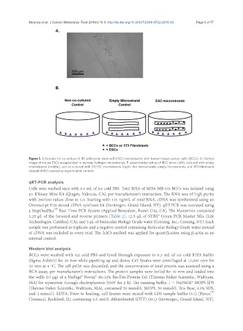
Figure 1. Schematic for co-culture of 3D embryonic stem cell (ESC)-microstrands with human breast cancer cells (BCCs). A: Optical
image of mouse ESCs encapsulated in alginate hydrogel microstrands; B: experimental set-up of BCC alone (left), cultured with empty
microstrands (middle), and co-cultured with 3D ESC-microstrands (right). No microstrands, empty microstrands, and 3T3 fibroblasts
instead of BCCs served as experimental controls
qRT-PCR analysis
Cells were washed once with 0.5 mL of ice cold PBS. Total RNA of MDA-MB-231 BCCs was isolated using
an RNeasy Mini Kit (Qiagen, Valencia, CA), per manufacturer’s instruction. The RNA was of high purity
with 260/280 values close to 2.0. Starting with 125 ng/mL of total RNA, cDNA was synthesized using an
Omniscript first-strand cDNA synthesis kit (Invitrogen, Grand Island, NY). qRT-PCR was executed using
TM
a StepOnePlus Real Time PCR System (Applied Biosystem, Foster City, CA). The Mastermix contained
1.25 µL of the forward and reverse primers [Table 2], 12.5 µL of SYBR® Green PCR Master Mix (Life
Technologies, Carlsbad, CA), and 5 µL of Molecular Biology Grade water (Corning, Inc., Corning, NY). Each
sample was performed in triplicate and a negative control containing Molecular Biology Grade water instead
of cDNA was included in every trial. The ∆∆Ct method was applied for quantification using β-actin as an
internal control.
Western blot analysis
BCCs were washed with ice cold PBS and lysed through exposure to 0.2 mL of ice cold RIPA buffer
(Sigma Aldrich) for 20 min while pipetting up and down. Cell lysates were centrifuged at 12,000 rpm for
20 min at 4 ºC. The cell pellet was discarded, and the concentration of total protein was assessed using a
BCA assay, per manufacturer’s instructions. The protein samples were boiled for 10 min and loaded into
the wells (15 µg) of a NuPage® Novex® 4%-12% Bis-Tris Protein Gel (Thermo Fisher Scientific, Waltham,
MA) for separation through electrophoresis (100V for 2 h). The running buffer, 1 × NuPAGE® MOPS SDS
(Thermo Fisher Scientific, Waltham, MA), contained 50 mmol/L MOPS, 50 mmol/L Tris Base, 0.1% SDS,
TM
and 1 mmol/L EDTA. Prior to loading, cell lysates were mixed with LDS sample buffer (4:1) (Pierce
Chemical, Rockford, IL) containing 0.5 mol/L dithiothreitol (DTT) (10:1) (Invitrogen, Grand Island, NY).