
Page 156 - Read Online
P. 156
Page 4 of 17 Mooney et al. J Cancer Metastasis Treat 2019;5:19 I http://dx.doi.org/10.20517/2394-4722.2018.93
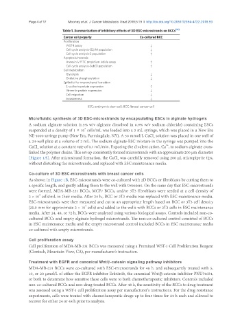
Table 1. Summarization of inhibitory effects of 3D ESC-microstrands on BCCs [10]
Cancer cell property Co-cultured BCC
Proliferation
WST-1 assay ↓
Cell cycle analysis-G2/M population ↓
Cell cycle analysis-S population ↑
Apoptosis/necrosis
Annexin-V FITC propidium iodide assay ↑
Cell cycle analysis-SubG1 population ↑
Cell metabolism
Glycolysis ↓
Oxidative phosphorylation ↓
Epithelial-to-mesenchymal transition
E-cadherin protein expression ↑
Vimentin protein expression ↓
Cell migration ↓
Invasiveness ↓
ESC: embryonic stem cell; BCC: breast cancer cell
Microfluidic synthesis of 3D ESC-microstrands by encapsulating ESCs in alginate hydrogels
A sodium alginate solution (1.5% w/v alginate dissolved in 0.9% w/v sodium chloride) containing ESCs
6
suspended at a density of 1 × 10 cells/mL was loaded into a 3 mL syringe, which was placed in a New Era
NE-1000 syringe pump (New Era, Farmingdale, NY). A 50 mmol/L CaCl solution was placed in one well of
2
a 24-well plate at a volume of 2 mL. The sodium alginate-ESC mixture in the syringe was pumped into the
2+
CaCl solution at a constant rate of 0.1 mL/min. Exposing the divalent cation, Ca , to sodium alginate cross-
2
linked the polymer chains. This set-up consistently formed microstrands with an approximate 200 µm diameter
[Figure 1A]. After microstrand formation, the CaCl was carefully removed using 200 µL micropipette tips,
2
without disturbing the microstrands, and replaced with ESC maintenance media.
Co-culture of 3D ESC-microstrands with breast cancer cells
As shown in Figure 1B, ESC-microstrands were co-cultured with 2D BCCs or fibroblasts by cutting them to
a specific length, and gently adding them to the well with tweezers. On the same day that ESC-microstrands
were formed, MDA-MB-231 BCCs, MCF7 BCCs, and/or 3T3 fibroblasts were seeded at a cell density of
4
2 × 10 cells/mL in their media. After 24 h., BCC or 3T3 media was replaced with ESC maintenance media.
ESC-microstrands were then measured and cut to an appropriate length based on BCC or 3T3 cell density
4
(35.0 mm for approximate 2 × 10 cells) and added to the wells with BCCs or 3T3 cells in ESC maintenance
media. After 24, 48, or 72 h, BCCs were analyzed using various biological assays. Controls included non-co-
cultured BCCs and empty alginate hydrogel microstrands. The non-co-cultured control consisted of BCCs
in ESC maintenance media and the empty microstrand control included BCCs in ESC maintenance media
co-cultured with empty microstrands.
Cell proliferation assay
Cell proliferation of MDA-MB-231 BCCs was measured using a Premixed WST-1 Cell Proliferation Reagent
(Clontech, Mountain View, CA), per manufacturer’s instruction.
Treatment with EGFR and canonical Wnt/β-catenin signaling pathway inhibitors
MDA-MB-231 BCCs were co-cultured with ESC-microstrands for 48 h. and subsequently treated with 5,
10, or 20 μmol/L of either the EGFR inhibitor Erlotinib, the canonical Wnt/β-catenin inhibitor PNU74654,
or both to determine how sensitive these cells were to both chemotherapeutic inhibitors. Controls included
non-co-cultured BCCs and non-drug-treated BCCs. After 48 h, the sensitivity of the BCCs to drug treatment
was assessed using a WST-1 cell proliferation assay per manufacturer’s instructions. For the drug resistance
experiments, cells were treated with chemotherapeutic drugs up to four times for 24 h each and allowed to
recover for either 24 or 48 h prior to analysis.