
Page 63 - Read Online
P. 63
Page 6 of 11 Narayanan et al. J Cancer Metastasis Treat 2019;5:36 I http://dx.doi.org/10.20517/2394-4722.2018.77
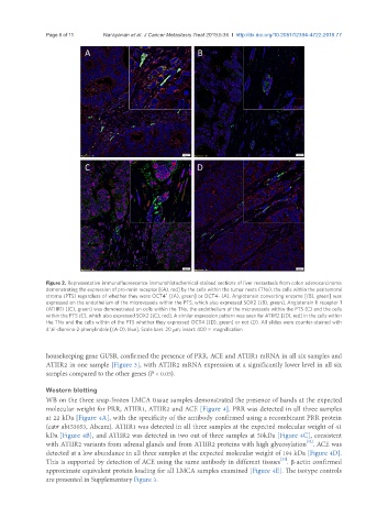
Figure 2. Representative immunofluorescence immunohistochemical-stained sections of liver metastasis from colon adenocarcinoma
demonstrating the expression of pro-renin receptor [(A), red] by the cells within the tumor nests (TNs), the cells within the peritumoral
+
stroma (PTS) regardless of whether they were OCT4 [(A), green] or OCT4- (A). Angiotensin converting enzyme [(B), green] was
expressed on the endothelium of the microvessels within the PTS, which also expressed SOX2 [(B), green]. Angiotensin II receptor 1
(ATIIR1) [(C), green] was demonstrated on cells within the TNs, the endothelium of the microvessels within the PTS (C) and the cells
within the PTS (C), which also expressed SOX2 [(C), red]. A similar expression pattern was seen for ATIIR2 [(D), red] in the cells within
the TNs and the cells within of the PTS whether they expressed OCT4 [(D), green] or not (D). All slides were counter-stained with
4’,6’-diamino-2-phenylindole [(A-D), blue]. Scale bars: 20 μm; insert: 400 × magnification
housekeeping gene GUSB, confirmed the presence of PRR, ACE and ATIIR1 mRNA in all six samples and
ATIIR2 in one sample [Figure 3], with ATIIR2 mRNA expression at a significantly lower level in all six
samples compared to the other genes (P < 0.05).
Western blotting
WB on the three snap-frozen LMCA tissue samples demonstrated the presence of bands at the expected
molecular weight for PRR, ATIIR1, ATIIR2 and ACE [Figure 4]. PRR was detected in all three samples
at 22 kDa [Figure 4A], with the specificity of the antibody confirmed using a recombinant PRR protein
(cat# ab153053, Abcam). ATIIR1 was detected in all three samples at the expected molecular weight of 41
kDa [Figure 4B], and ATIIR2 was detected in two out of three samples at 50kDa [Figure 4C], consistent
[32]
with ATIIR2 variants from adrenal glands and from ATIIR2 proteins with high glycosylation . ACE was
detected at a low abundance in all three samples at the expected molecular weight of 194 kDa [Figure 4D].
[33]
This is supported by detection of ACE using the same antibody in different tissues . β-actin confirmed
approximate equivalent protein loading for all LMCA samples examined [Figure 4E]. The isotype controls
are presented in Supplementary Figure 3.