
Page 648 - Read Online
P. 648
John et al. Hepatoma Res 2020;6:56 I http://dx.doi.org/10.20517/2394-5079.2020.37 Page 7 of 9
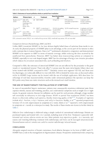
Table 3. Outcomes of recent palliative whole or partial liver irradiation
Dose/fractionation
Authors Primary tumor n Treatment Outcome Toxicities
(#)
Bydder et al. [33] CRC 39% 28 WLRT/PLRT 10 Gy/2 # 54% partial or 2 patients with
2003 NSCLC 4% complete symptomatic Grade 3 vomiting
Esophageal 11% response and diarrhoea
SCLC 7%
Other 29%
Yin et al. [35] CRC 19 WLRT + tumor boost + 53.4 Gy (including 52.6% overall response 2 patients with Grade
2014 concurrent chemotherapy boost)/# NR 3 elevated bilirubin
Edyta et al. [34] Colon 59% 27 WLRT Mean 17 Gy/ 40% partial or 1 patient with Grade
2015 Stomach 26% 5-12 # complete symptomatic 3 vomiting and
Pancreas 15% response diarrhoea
CRC: colorectal cancer; NSCLC: non-small cell lung cancer; SCLC: small cell lung cancer; NR: not reported
Comparison between Brachytherapy, SBRT and RFA
Unlike SBRT, interstitial HDRBT to the liver delivers highly lethal doses of radiation from inside to out.
As such, the physical property of HDRBT plays to its advantage, as the central part of the tumour is often
[29]
radio-resistant due to tumour hypoxia. Hass et al. performed a dosimetric comparison and demonstrated
HDRBT to be superior to SBRT in terms of tumour coverage, whilst reducing the dose received by the
unaffected liver parenchyma. However, SBRT has better dose manoeuvring options as compared to
HDRBT, especially in non-oval shaped targets. SBRT has the advantage of being a non-invasive procedure
which reduces the procedure-associated risks, such as bleeding and infection.
Compared to RFA, the outcomes of interstitial HDRBT liver are not affected by the proximity to the great
vessels or vascularized tumour (“heat sink effect”). Lesions near the main intra-hepatic biliary ducts are
[22]
better treated with HDRBT compared to RFA . Similarly, lesions near segment VII/VIII, and those near
the diaphragm, are technically difficult to treat with RFA. RFA is limited by lesion size, as discussed earlier,
whilst in HDRBT, large lesions can be treated with the use of multiple applicators. RFA does have the
advantage of real-time manoeuvrability, whereby under ultrasound guidance the operator can keep the
RFA probe away from non-static structures such as the stomach and small intestine.
THE USE OF RADIOTHERAPY FOR PALLIATION OF SYMPTOMS
In cases of uncontrolled hepatic metastases, patients may consequently experience abdominal pain (from
capsular stretch), nausea and vomiting, jaundice, and constitutional symptoms such as weight loss or night
sweats. In general, systemic therapy for palliation can be used for such patients, although a large number
will eventually be refractory in end-stage disease. WLRT or partial liver irradiation (PLRT) has been
shown to effectively palliate such patients, thereby improving quality of life [30-32] . Examples of regimens
include 8Gy/1 fraction, 21Gy/7 fractions, and 30Gy/15 fractions. Bydder et al. reported prospectively
[33]
[34]
between 53% to 66% improvement in symptoms at 2 weeks. Edyta et al. reported a 100% improvement
in symptoms at 1 month in a retrospective study. The results of these studies are shown in further detail in
Table 3.
Palliative liver radiotherapy is delivered using a simple method of conventional radiotherapy. Patients are
positioned supine and treated with 2 to 3 portals, including most of the liver. Treatment is generally well-
tolerated and serious adverse events are rare. Most patients may experience grade 1 to 2 anorexia, and
nausea and vomiting following treatment with radiotherapy, and these can be managed symptomatically.
Dexamethasone and anti-emetics are useful to counter radiotherapy-induced nausea.
CONCLUSION
Alongside surgical resection of hepatic metastases, local ablative therapies in the form of SBRT and CT-
HDRBT have a role in the management of oligometastatic disease. Prospective randomized trials comparing