
Page 646 - Read Online
P. 646
John et al. Hepatoma Res 2020;6:56 I http://dx.doi.org/10.20517/2394-5079.2020.37 Page 5 of 9
The role of interstitial brachytherapy for oligometastatic liver disease
Technique
Although brachytherapy has a long history in oncology, it was not until the early 1980s when it was used
[17]
for liver tumours. Dritschilo et al. described the percutaneous implantation of interstitial brachytherapy
applicators under sonographic guidance in 1986. Subsequently, intraoperative catheter placement (under
[18]
direct visualisation and/or sonography-assisted) has also been described . Later, in 2004, Ricke et al.
[19]
published a phase II trial using CT-guidance for interstitial high dose-rate brachytherapy (HDRBT) of liver
tumours which were unsuitable for thermal ablation.
The choice of image guidance for interstitial liver HDRBT application is operator-specific. In general,
the use of CT, or a hybrid method combining ultrasound and CT, is the preferred choice. CT guidance
allows better visualization of gastrointestinal structures, major vessels and intrahepatic bile ducts. The use
of ultrasound, even when combined with CT, can reduce radiation exposure of the healthcare personnel
involved.
The procedure is performed under sterile conditions using local anaesthesia and mild to moderate sedation.
The skin puncture site is identified based on CT images. Under CT-guidance, the applicator is advanced
past the liver capsule, into the target, in the same phase of breathing. The applicators are advanced at least
5 mm beyond the target, to account for breathing motion. After insertion of the applicators, CT or MRI
with contrast is usually performed with thin axial cuts (2 mm) for applicator reconstruction. Doses in the
range of 15-25 Gy in a single fraction are usually prescribed depending on the histology and organs-at-risk
tolerance. Colorectal and sarcoma metastases tend to be radioresistant, and are usually treated with 25 Gy.
[20]
In view of the clear dose-response reported by Ricke et al. , higher doses should be used when possible.
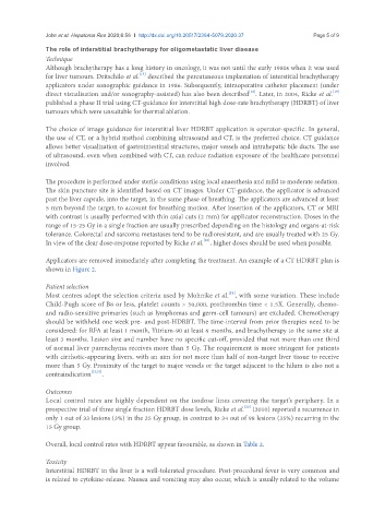
Applicators are removed immediately after completing the treatment. An example of a CT HDRBT plan is
shown in Figure 2.
Patient selection
[21]
Most centres adopt the selection criteria used by Mohnike et al. , with some variation. These include
Child-Pugh score of B8 or less, platelet counts > 50,000, prothrombin time < 1.5X. Generally, chemo-
and radio-sensitive primaries (such as lymphomas and germ-cell tumours) are excluded. Chemotherapy
should be withheld one week pre- and post-HDRBT. The time-interval from prior therapies need to be
considered: for RFA at least 1 month, Yttrium-90 at least 6 months, and brachytherapy to the same site at
least 3 months. Lesion size and number have no specific cut-off, provided that not more than one third
of normal liver parenchyma receives more than 5 Gy. The requirement is more stringent for patients
with cirrhotic-appearing livers, with an aim for not more than half of non-target liver tissue to receive
more than 5 Gy. Proximity of the target to major vessels or the target adjacent to the hilum is also not a
contraindication [22,23] .
Outcomes
Local control rates are highly dependent on the isodose lines covering the target’s periphery. In a
[20]
prospective trial of three single fraction HDRBT dose levels, Ricke et al. (2010) reported a recurrence in
only 1 out of 33 lesions (3%) in the 25 Gy group, in contrast to 34 out of 98 lesions (35%) recurring in the
15 Gy group.
Overall, local control rates with HDRBT appear favourable, as shown in Table 2.
Toxicity
Interstitial HDRBT in the liver is a well-tolerated procedure. Post-procedural fever is very common and
is related to cytokine-release. Nausea and vomiting may also occur, which is usually related to the volume