
Page 244 - Read Online
P. 244
Page 2 of 8 Dutta et al. Hepatoma Res 2019;5:23 I http://dx.doi.org/10.20517/2394-5079.2019.09
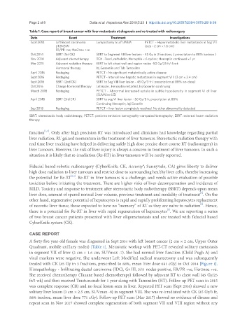
Table 1. Case report of breast cancer with liver metastasis at diagnosis and re-treated with radiosurgery
Date Event Treatment Investigations
Sept 2014 Left Breast carcinoma Lumpectomy à Left MRM PETCT – Hypermetabolic liver metastases in Seg VII
pT2N2M1 (size - 2 cm × 1.6 cm)
ER/PR +ve; Her2neu +ve
Oct 2014 SBRT (1st CK) SBRT to Segment VII liver lesions – 45 Gy in 3 fractions, ( prescription to 88% isodose )
Nov 2014 Adjuvant chemotherapy TCH –Taxol, carbolatin, Herceptin × 6 cycles ; Herceptin continued × 1 yr
Mar 2015 Adjuvant radiation therapy EBRT to left chest wall and region nodes -50 Gy/25 fr/ 5 wk
Hormonal therapy Inj Goserelin and Tab Tamoxifen
April 2016 Restaging PETCT - No significant metabolically active disease
Sept 2016 Restaging PETCT - Interval new hepatic metastases in segment VIII (3 cm × 2.4 cm)
Sept 2016 SBRT (2nd CK) SBRT to Seg VIII liver lesion – 45 Gy/3 fr ( prescription at 88% iso-dose)
Oct 2016 Change hormonal therapy Letrazole , Herceptin restarted, Inj Goserlin continuing
March 2018 Restaging PETCT - Abnormal increased uptake in subtle hypodensity in segment VI of liver
(SUVMax 6.5)
April 2018 SBRT (3rd CK) SBRT to seg VI liver lesion - 50 Gy/5 fr, prescription at 88%
Continuing Herceptin, Inj Goserlin
Sep 2018 Restaging PETCT – liver lesion completely resolved. No other abnormality detected
SBRT: stereotactic body radiotherapy; PETCT: positron emission tomography–computed tomography; EBRT: external beam radiation
therapy
[1,2]
function . Only after high precision RT was introduced and clinicians had knowledge regarding partial
liver radiation, RT gained momentum in the treatment of liver tumours. Stereotactic radiation therapy with
real time liver tracking have helped in delivering safely high dose precise short course RT (radiosurgery) in
liver tumours. However, the risk of liver injury is always a concern in treatment of liver tumours. In such a
situation it is likely that re-irradiation (Re-RT) in liver tumours will be rarely reported.
Fiducial based robotic radiosurgery (CyberKnife, CK, Accuray®; Sunnyvale, CA) gives liberty to deliver
high dose radiation to liver tumours and restrict dose to surrounding healthy liver cells, thereby increasing
the potential for Re-RT . Re-RT in liver tumours is a challenge, and needs active evaluation of possible
[3,4]
toxicities before initiating the treatment. There are higher risks of liver decompensation and incidence of
RILD. Toxicity and response to treatment after stereotactic body radiotherapy (SBRT) depends upon mean
[5]
liver dose, amount of spared normal liver volume, previous treatment and modality of treatment . On the
other hand, regenerative potential of hepatocytes is rapid and rapidly proliferating hepatocytes replacement
[2]
of necrotic liver tissue, those expected to have no “memory” of RT as they are naïve to radiation . Hence,
there is a potential for Re-RT in liver with rapid regeneration of hepatocytes . We are reporting a series
[2]
of two breast cancer patients presented with liver oligometastasis and are treated with fiducial based
CyberKnife system (CK).
CASE REPORT
A forty-five year-old female was diagnosed in Sept 2014 with left breast cancer (2 cm × 2 cm, Upper Outer
Quadrant, mobile axillary nodes) [Table 1]. Metastatic workup with PET-CT revealed solitary metastasis
in segment VII of liver (2 cm × 1.6 cm, SUVmax -7). She had normal liver function (Child Pugh A) and
viral markers were negative. She underwent Left Modified radical mastectomy and was subsequently
treated with CK (45 Gy in 3 fractions, prescribed to 88%, mean liver dose 681 cGy) in Oct 2014 [Figure 1].
Histopathology - Infiltrating ductal carcinoma (IDC), Gr III, 5/11 nodes positive, ER/PR +ve, Her2neu +ve.
She received chemotherapy (Taxane based chemotherapy) followed by adjuvant RT to chest wall (45 Gy/25
fr/5 wk) and then received Trastuzumab for 1 year along with Tamoxifen (HT). Follow up PET scan in 2015
was complete response (CR) and no focal lesion seen in liver. Repeated PET scan (Sept 2016) showed a new
solitary liver lesion (3 cm × 2.5 cm, SUVmax -8) in segment VIII. She was re-irradiated with CK (45 Gy/3 fr,
88% isodose, mean liver dose 771 cGy). Follow-up PET scan (Mar 2017) showed no evidence of disease and
repeat scan in Nov 2017 showed complete regeneration of both segment VII and VIII region without any