
Page 248 - Read Online
P. 248
Page 6 of 8 Dutta et al. Hepatoma Res 2019;5:23 I http://dx.doi.org/10.20517/2394-5079.2019.09
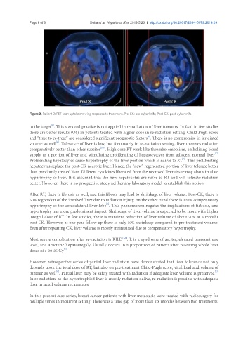
Figure 3. Patient 2: PET scan uptake showing response to treatment. Pre-CK: pre-cyberknife; Post-CK: post-cyberknife
[9]
to the target . This standard practice is not applied in re-radiation of liver tumours. In fact, in few studies
there are better results (OS) in patients treated with higher dose in re-radiation setting. Child Pugh Score
[6]
and “time to re-treat” are considered significant prognostic factors . There is no compromise in irridiated
[6]
volume as well . Tolerance of liver is low, but fortunately in re-radiation setting, liver tolerates radiation
[2,5]
comparatively better than other subsites . High dose RT work like thrombo-embolism, embolizing blood
[7]
supply to a portion of liver and stimulating proliferating of hepatocytcyes from adjacent normal liver .
Proliferating hepatocytes cause hypertrophy of the liver portion which is naiive to RT . This proliferating
[7]
hepatocytes replace the post-CK necrotic liver. Hence, the “new” regenerated portion of liver tolerate better
than previously treated liver. Different cytokines liberated from the necrosed liver tissue may also stimulate
hypertrophy of liver. It is assumed that the new hepatocytes are naïve to RT and will tolerate radiation
better. However, there is no prospective study neither any laboratory model to establish this notion.
After RT, there is fibrosis as well, and this fibosis may lead to shrinkage of liver volume. Post-CK, there is
50% regression of the involved liver due to radiation injury, on the other hand there is 320% compensatory
[2]
hypertrophy of the contralateral liver lobe . This phenomenon negates the implications of firbosis, and
hypertrophy has more predominent impact. Shrinkage of liver volume is expected to be more with higher
integral dose of RT. In few studies, there is transient reduction of liver volume of about 20% at 3 months
post-CK. However, at one year follow up there is only 10% shrinkage compared to pre-treatment volume.
Even after repeating CK, liver volume is mostly maintained due to compensatory hypertrophy.
[7,8]
Most severe complication after re-radiation is RILD . It is a syndrome of ascites, elevated transaminase
level, and anicteric hepatomegaly. Usually occurs in a proportion of patient after receiving whole liver
[8]
doses of > 30-35 Gy .
However, retrospective series of partial liver radiation have demonstrated that liver tolerance not only
depends upon the total dose of RT, but also on pre-treatment Child-Pugh score, viral load and volume of
[3]
[8]
tumour as well . Partial liver may be safely treated with radiation if adequate liver volume is preserved .
In re-radiation, as the hypertrophied liver is mostly radiation naiive, re-radiation is possible with adequate
dose in small volume recurrences.
In this present case series, breast cancer patients with liver metastasis were treated with radiosurgery for
multiple times in recurrent setting. There was a time gap of more than six months between two treatments.