
Page 246 - Read Online
P. 246
Page 4 of 8 Dutta et al. Hepatoma Res 2019;5:23 I http://dx.doi.org/10.20517/2394-5079.2019.09
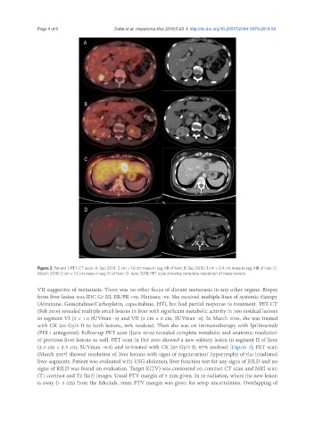
Figure 2. Patient 1: PET-CT scan. A: Sep 2014: 2 cm × 1.6 cm mass in seg VII of liver; B: Sep 2016: 3 cm × 2.4 cm mass in seg VIII of liver; C:
March 2018: 2 cm × 1.5 cm mass in seg VI of liver; D: June 2018: PET scan showing complete resolution of mass lesions
VII suggestive of metastasis. There was no other focus of distant metastasis in any other organs. Biopsy
from liver lesion was IDC Gr III, ER/PR +ve, Her2neu -ve. She received multiple lines of systemic therapy
(Abraxane, Gemcitabine/Carboplatin, capecitabine, HT), but had partial response to treatment. PET-CT
(Feb 2016) revealed multiple small lesions in liver with significant metabolic activity in two residual lesions
in segment VI (2 × 1.5 SUVmax -5) and VII (2 cm × 2 cm, SUVmax -8). In March 2016, she was treated
with CK (45 Gy/3 fr to both lesions, 86% isodose). Then she was on immunotherapy with Ipilimumab
(PDL1 antagonist). Follow-up PET scan (June 2016) revealed complete metabolic and anatomic resolution
of previous liver lesions as well. PET scan in Oct 2016 showed a new solitary lesion in segment II of liver
(2.3 cm × 2.5 cm, SUVmax -8.0) and re-treated with CK (45 Gy/3 fr, 87% isodose) [Figure 3]. PET scan
(March 2017) showed resolution of liver lesions with signs of regeneration/ hypertrophy of the irradiated
liver segments. Patient was evaluated with USG abdomen, liver function test for any signs of RILD and no
signs of RILD was found on evaluation. Target (GTV) was contoured on contrast CT scan and MRI scan
(T1 contrast and T2 flair) images. Usual PTV margin of 3 mm given. In re-radiation, where the new lesion
is away (> 5 cm) from the fiducials, 5mm PTV margin was given for setup uncertainties. Overlapping of