
Page 239 - Read Online
P. 239
Page 6 of 8 Block et al. Hepatoma Res 2019;5:21 I http://dx.doi.org/10.20517/2394-5079.2019.17
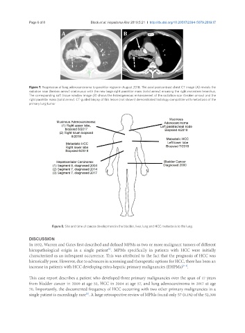
Figure 7. Progression of lung adenocarcinoma to parahilar region in August 2018. The axial postcontrast chest CT image (A) reveals the
radiation scar (broken arrow) continuous with the new large right parahilar mass (solid arrow) encasing the right mainstem bronchus;
The corresponding soft tissue window image (B) shows the heterogeneous enhancement of the radiation scar (broken arrow) and the
right parahilar mass (solid arrow). CT-guided biopsy of this lesion (not shown) demonstrated histology compatible with metastasis of the
primary lung tumor
Figure 8. Site and time of cancer development in the bladder, liver, lung and HCC metastasis to the lung.
DISCUSSION
In 1932, Warren and Gates first described and defined MPMs as two or more malignant tumors of different
histopathological origin in a single patient . MPMs specifically in patients with HCC were initially
[1]
characterized as an infrequent occurrence. This was attributed to the fact that the prognosis of HCC was
historically poor. However, due to advances in screening and therapeutic options for HCC, there has been an
increase in patients with HCC developing extra-hepatic primary malignancies (EHPMs) .
[2-4]
This case report describes a patient who developed three primary malignancies over the span of 17 years
from bladder cancer in 2000 at age 53, HCC in 2004 at age 57, and lung adenocarcinoma in 2017 at age
70. Importantly, the documented frequency of HCC occurring with two other primary malignancies in a
single patient is exceedingly rare . A large retrospective review of MPMs found only 57 (0.1%) of the 52,398
[2]