
Page 237 - Read Online
P. 237
Page 4 of 8 Block et al. Hepatoma Res 2019;5:21 I http://dx.doi.org/10.20517/2394-5079.2019.17
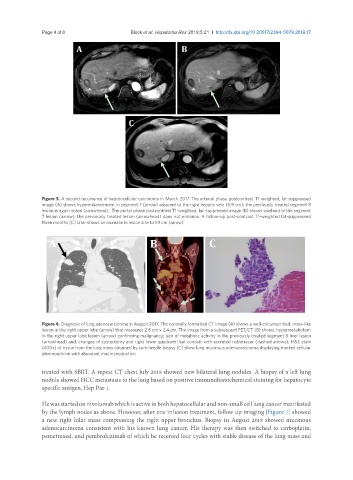
Figure 3. A second recurrence of hepatocellular carcinoma in March 2017. The arterial phase postcontrast T1-weighted, fat-suppressed
image (A) shows hyperenhancement in segment 7 (arrow) adjacent to the right hepatic vein (0.9 cm); the previously treated segment 8
lesion is again noted (arrowhead). The portal phase postcontrast T1-weighted, fat-suppressed image (B) shows washout in the segment
7 lesion (arrow); the previously treated lesion (arrowhead) does not enhance. A follow-up post-contrast T1-weighted fat-suppressed
three months (C) later shows an increase in lesion size to 1.9 cm (arrow)
Figure 4. Diagnosis of lung adenocarcinoma in August 2017. The coronally formatted CT image (A) shows a well-circumscribed, mass-like
lesion in the right upper lobe (arrow) that measures 2.8 cm × 2.4 cm; The image from a subsequent PET/CT (B) shows: hypermetabolism
in the right upper lobe lesion (arrow) confirming malignancy; lack of metabolic activity in the previously treated segment 8 liver lesion
(arrowhead) and; changes of cystectomy and right lower quadrant ileal conduit with excreted radiotracer (dashed arrows); H&E stain
(400x) of tissue from the lung mass obtained by core needle biopsy (C) show lung mucinous adenocarcinoma displaying marked cellular
pleomorphism with abundant mucin production
treated with SBRT. A repeat CT chest July 2018 showed new bilateral lung nodules. A biopsy of a left lung
nodule showed HCC metastasis to the lung based on positive immunohistochemical staining for hepatocyte
specific antigen, Hep Par-1.
He was started on nivolumab which is active in both hepatocellular and non-small cell lung cancer manifested
by the lymph nodes as above. However, after one infusion treatment, follow-up imaging [Figure 7] showed
a new right hilar mass compressing the right upper bronchus. Biopsy in August 2018 showed mucinous
adenocarcinoma consistent with his known lung cancer. His therapy was then switched to carboplatin,
pemetrexed, and pembroluzimab of which he received four cycles with stable disease of the lung mass and