
Page 77 - Read Online
P. 77
Mehta et al. Hepatoma Res 2018;4:7 I http://dx.doi.org/10.20517/2394-5079.2017.35 Page 3 of 9
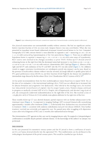
Figure 1. Hyper attenuating lesion from prior conventional transarterial chemoembolization containing lipiodol (white arrow)
Her physical examination was unremarkable notably without asterixis. She had no significant cardiac
history (ejection fraction of 55% on stress test). Surgical history was non contributive. When she was
presented in transplant tumor board, abdominal ultrasound showed several lesions in the liver. Computed
tomography (CT) with contrast showed a mass identified in segments 6 and 7 measuring 4.6 cm × 4.0 cm
× 4.5 cm with surrounding hypoattenuation of the liver parenchyma [Figure 1]. There was an additional
hypodense lesion in segment 2 measuring 3.1 cm × 4.9 cm × 3.5 cm with some areas of hyperdensity.
Both lesions were deemed to be changes secondary to prior TACE. Follow-up CT showed arterial
enhancing lesions in the right liver lobe the dominant lesion had increased in size from 4.6 cm × 4.0 cm ×
4.5 cm to 9.2 cm × 8.9 cm × 11.0 cm with washout, characteristics HCC findings [Figure 2]. Absence of the
right and left PV and confluence of the SV and SMV into the IVC was also noted [Figure 3]. No collateral
vessels to suggest cavernous transformation nor extrahepatic portal vein remnant can be seen. Based on
the presence of multinodular disease without vascular invasion (although difficult to qualify given lack of
PV), good performance status (ECOG 0), and liver function (Child Pugh B), her disease was classified as
[7]
intermediate stage disease by the Barcelona Clinic Liver Classification (BCLC) system or BCLC B .
At that point recommendation from the liver multidisciplinary tumor board was to repeat TACE. Yet, at
the time of her evaluation in the interventional radiology clinic, TACE was not offered due to increased risk
for abscess formation and progressive liver dysfunction. After referral to the oncology team, she received
two intra-arterial chemoinfusions of cisplatin into the proper hepatic artery. Patient’s disease continued
to progress as markedly elevated AFP of 8779. Despite risk of hepatoxicity and elevated lung shunt of
21%, she subsequently underwent radioembolization to the right lobe of the liver. She received a dose of
1.06 gBq (29.1 mCi) of Yttrium-90 (Y-90) embolic resin spheres delivered to the right lobe of the liver.
Three months follow-up CT scan showed dramatic partial response with no further enhancement in the
dominant mass [Figure 4]. Incongruent to imaging findings AFP increased dramatically normalizing
approximately 9 months after treatment [Table 1]. Unfortunately liver dysfunction was exacerbated due
to treatment [Table 1] consistent with radiation embolization induced liver disease (REILD). Only further
treatment received was octreotide and supportive care. She passed away after struggling with depression
8 years after initial diagnosis of HCC and 22 months after radioembolization therapy.
The determination of PV agenesis in this case was by imaging features only. No surgical or histopathological
confirmation is available despite patient’s ultimate demise. To the knowledge of the authors’ no autopsy was
performed.
DISCUSSION
In the case presented the mesenteric venous system and the SV joined to form a confluence of vessels
yet this confluence drained directly into the suprarenal IVC. This malformation can be attributed to the
embryological development of the portal venous system. This would be classified as type 1b shunt.