
Page 80 - Read Online
P. 80
Page 6 of 9 Mehta et al. Hepatoma Res 2018;4:7 I http://dx.doi.org/10.20517/2394-5079.2017.35
A B
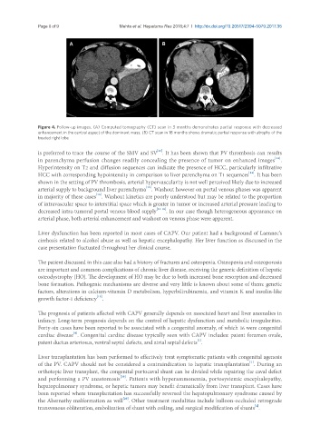
Figure 4. Follow-up images. (A) Computed tomography (CT) scan in 3 months demonstrates partial response with decreased
enhancement in the central aspect of the dominant mass; (B) CT scan in 18 months shows dramatic partial response with atrophy of the
treated right lobe
[13]
is preferred to trace the course of the SMV and SV . It has been shown that PV thrombosis can results
[14]
in parenchyma perfusion changes readily concealing the presence of tumor on enhanced images .
Hyperintensity on T2 and diffusion sequences can indicate the presence of HCC, particularly infiltrative
[14]
HCC with corresponding hypointensity in comparison to liver parenchyma on T1 sequences . It has been
shown in the setting of PV thrombosis, arterial hypervascularity is not well perceived likely due to increased
[15]
arterial supply to background liver parenchyma . Washout however on portal venous phases was apparent
[15]
in majority of these cases . Washout kinetics are poorly understood but may be related to the proportion
of intravascular space to interstitial space which is greater in tumor or increased arterial pressure leading to
decreased intra-tumoral portal venous blood supply [15-18] . In our case though heterogeneous appearance on
arterial phase, both arterial enhancement and washout on venous phase were apparent.
Liver dysfunction has been reported in most cases of CAPV. Our patient had a background of Laennec’s
cirrhosis related to alcohol abuse as well as hepatic encephalopathy. Her liver function as discussed in the
case presentation fluctuated throughout her clinical course.
The patient discussed in this case also had a history of fractures and osteopenia. Osteopenia and osteoporosis
are important and common complications of chronic liver disease, receiving the generic definition of hepatic
osteodystrophy (HO). The development of HO may be due to both increased bone resorption and decreased
bone formation. Pathogenic mechanisms are diverse and very little is known about some of them: genetic
factors, alterations in calcium-vitamin D metabolism, hyperbilirubinemia, and vitamin K and insulin-like
[19]
growth factor-1 deficiency .
The prognosis of patients affected with CAPV generally depends on associated heart and liver anomalies in
infancy. Long-term prognosis depends on the control of hepatic dysfunction and metabolic irregularities.
Forty-six cases have been reported to be associated with a congenital anomaly, of which 16 were congenital
[4]
cardiac disease . Congenital cardiac disease typically seen with CAPV includes: patent foramen ovale,
[1]
patent ductus arteriosus, ventral septal defects, and atrial septal defects .
Liver transplantation has been performed to effectively treat symptomatic patients with congenital agenesis
[1]
of the PV. CAPV should not be considered a contraindication to hepatic transplantation . During an
orthotopic liver transplant, the congenital portocaval shunt can be divided while repairing the caval defect
[20]
and performing a PV anastomosis . Patients with hyperammonemia, portosystemic encephalopathy,
hepatopulmonary syndrome, or hepatic tumors may benefit dramatically from liver transplant. Cases have
been reported where transplantation has successfully reversed the hepatopulmonary syndrome caused by
[20]
the Abernathy malformation as well . Other treatment modalities include balloon-occluded retrograde
[4]
transvenous obliteration, embolization of shunt with coiling, and surgical modification of shunts .