
Page 78 - Read Online
P. 78
Page 4 of 9 Mehta et al. Hepatoma Res 2018;4:7 I http://dx.doi.org/10.20517/2394-5079.2017.35
A B
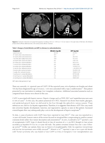
Figure 2. Contrast enhanced computed tomography revealing a 9.2 cm × 8.9 cm × 11.0 cm mass in the right lobe of the liver. (A) Arterial
phase shows diffuse enhancement; (B) venous phase shows washout
Table 1. Changes of total bilirubin and AFP in reference to radioembolization
Component Bilirubin (mg/dL) AFP (ng/mL)
Latest reference 0.0-1.2 0.0-8.3
21 months after radioembolization 12.7 6.7
12 months after radioembolization 13.9 20.7 (H)
11 months after radioembolization 13.8 55.3 (H)
10 months after radioembolization 8.4 178.2 (H)
9 months after radioembolization 6.9 805.4 (H)
7 months after radioembolization 6.9 5450.0 (H)
6 months after radioembolization 7.4 19,394.0 (H)
4 months after radioembolization 7.4 > 60,500.0 (H)
3 months after radioembolization 4.8 55,658.0 (H)
2 weeks after radioembolization 2.9 18,662.0 (H)
1 month prior to radioembolization 2.1 8779.0 (H)
2 months prior to radioembolization 1.8 3678.0 (H)
4 months prior to radioembolization 1.6 1032.0 (H)
AFP: alpha fetal protein; H: high
There are currently 101 reported cases of CAPV. Of the reported cases, 66% of patients are females and about
[4]
70% had been diagnosed by age of 18 years; < 10% were associated with a type 2 malformation . This patient
presented to our institution to undergo liver transplant evaluation. Additional associated anomalies such as
congenital heart disease were absent in this case.
CAPV is associated with hepatic tumors. Hepatic changes such as FNH, HCC and hepatoblastoma were seen
[1]
in 40% of cases . In this case, the patient presented with HCC. Research has shown that insulin, glucagon,
and epidermal growth factor are delivered to the liver through the splanchnic venous system. These
substances are vital for the hepatic regeneration. Therefore, it is suggested that absence of PV flow may result
into abnormal hepatic development, function, and regenerative capacity as seen in this patient. Increased
[8]
arterial hepatic flow may subsequently play a role in the development of hepatic neoplasms .
[1]
To date, 4 cases of patients with CAPV have been reported to have HCC . One case was reported in a
14-year-old female, however nature of the review focused on intestinal flora compensating to result in normal
[10]
[9]
ammonia levels rather than tumor description and presentation . In 2001, Lundstedt et al. eported a case
of asymptomatic CAPV (type 1b shunt) found at time of resection of a 12-cm HCC thought to have arisen
secondary to hepatitis B virus in a 51-year-old male. The patient remained disease free over 2-year follow-
[10]
up period Unlike our patient, there was no history of encephalopathy. Only the aspartate transaminase
[11]
[10]
and alanine transaminase were mildly elevated . Morotti et al. reported a case of an 8-year-old female
with Turner syndrome who was found to have CAPV at time of transplant. Liver transplantation was