
Page 79 - Read Online
P. 79
Mehta et al. Hepatoma Res 2018;4:7 I http://dx.doi.org/10.20517/2394-5079.2017.35 Page 5 of 9
A B
C D
E F
Confluence
Inferior
vena
cava Splenic vein
Superior mesenteric vein
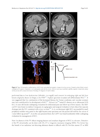
Figure 3. Type 1B Abernathy malformation. (A-D) Axial computed tomography images showing course of hepatic artery (black arrow),
confluence of superior mesenteric vein and splenic vein (white arrow), inferior vena cava (asterisk), and the superior mesenteric artery
(white arrow); (E) coronal reformats of findings; (F) diagram of malformation
performed due to liver dysfunction (bilirubin 13.9 mg/dL) and concern for enlarging right and left liver
[11]
[11]
lesions originally shown on biopsy to be FNH . Explant specimen revealed well-differentiated HCC . It
was suggested that the combination of the hormonal therapy for Turner syndrome, and vascular anomaly
[11]
[12]
may have contributed to the development of HCC . Pichon et al. noted PV absence on an ultrasound (US)
for a 36-year-old female undergoing evaluation for abdominal pain and follow-up of liver masses. The SMV
and SV were found on indirect venogram at angiography and surgical evaluation to have direct but separate
[12]
drainage into the IVC consistent with a type 1a shunt . A 12-cm dominant right HCC surrounded by small
peripheral nodules were noted in the right hepatectomy specimen and the patient did well for the course of
[12]
2-year follow-up period . As in the case presented here, the CAPV was found incidentally while undergoing
evaluation for management of HCC.
How the absence of the PV effects imaging features and resultant diagnosis of HCC is unknown. Detection
of the PV abnormality can be done with US, CT or magnetic resonance imaging (MRI). The former has
the benefit of no radiation, but detecting alternate shunts is difficult with US. For this reason CT or MRI