
Page 84 - Read Online
P. 84
Xiong et al. Nature of liver lymphoma
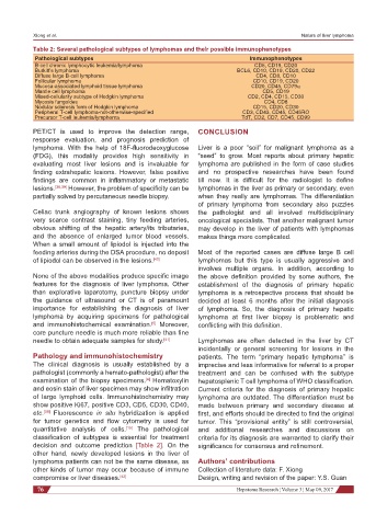
Table 2: Several pathological subtypes of lymphomas and their possible immunophenotypes
Pathological subtypes Immunophenotypes
B-cell chronic lymphocytic leukemia/lymphoma CD5, CD19, CD20
Burkitt’s lymphoma BCL6, CD10, CD19, CD20, CD22
Diffuse large B-cell lymphoma CD4, CD8, CD10
Follicular lymphoma CD10, CD19, CD20
Mucosa-associated lymphoid tissue lymphoma CD20, CD45, CD79α
Mantle cell lymphoma CD5, CD19
Mixed-cellularity subtype of Hodgkin lymphoma CD2, CD4, CD15, CD30
Mycosis fungoides CD4, CD8
Nodular sclerosis form of Hodgkin lymphoma CD15, CD20, CD30
Peripheral T-cell lymphoma-not-otherwise-specified CD3, CD43, CD45, CD45RO
Precursor T-cell leukemia/lymphoma TdT, CD2, CD7, CD45, CD99
PET/CT is used to improve the detection range, CONCLUSION
response evaluation, and prognosis prediction of
lymphoma. With the help of 18F-fluorodeoxyglucose Liver is a poor “soil” for malignant lymphoma as a
(FDG), this modality provides high sensitivity in “seed” to grow. Most reports about primary hepatic
evaluating most liver lesions and is invaluable for lymphoma are published in the form of case studies
finding extrahepatic lesions. However, false positive and no prospective researches have been found
findings are common in inflammatory or metastatic till now. It is difficult for the radiologist to define
lesions. [38,39] However, the problem of specificity can be lymphomas in the liver as primary or secondary, even
partially solved by percutaneous needle biopsy. when they really are lymphomas. The differentiation
of primary lymphoma from secondary also puzzles
Celiac trunk angiography of known lesions shows the pathologist and all involved multidisciplinary
very scarce contrast staining, tiny feeding arteries, oncological specialists. That another malignant tumor
obvious shifting of the hepatic artery/its tributaries, may develop in the liver of patients with lymphomas
and the absence of enlarged tumor blood vessels. makes things more complicated.
When a small amount of lipiodol is injected into the
feeding arteries during the DSA procedure, no deposit Most of the reported cases are diffuse large B cell
of lipiodol can be observed in the lesions. [40] lymphomas but this type is usually aggressive and
involves multiple organs. In addition, according to
None of the above modalities produce specific image the above definition provided by some authors, the
features for the diagnosis of liver lymphoma. Other establishment of the diagnosis of primary hepatic
than explorative laparotomy, puncture biopsy under lymphoma is a retrospective process that should be
the guidance of ultrasound or CT is of paramount decided at least 6 months after the initial diagnosis
importance for establishing the diagnosis of liver of lymphoma. So, the diagnosis of primary hepatic
lymphoma by acquiring specimens for pathological lymphoma at first liver biopsy is problematic and
and immunohistochemical examination. Moreover, conflicting with this definition.
[5]
core puncture needle is much more reliable than fine
needle to obtain adequate samples for study. [41] Lymphomas are often detected in the liver by CT
incidentally or general screening for lesions in the
Pathology and immunohistochemistry patients. The term “primary hepatic lymphoma” is
The clinical diagnosis is usually established by a imprecise and less informative for referral to a proper
pathologist (commonly a hemato-pathologist) after the treatment and can be confused with the subtype
examination of the biopsy specimens. Hematoxylin hepatosplenic T cell lymphoma of WHO classification.
[4]
and eosin stain of liver specimen may show infiltration Current criteria for the diagnosis of primary hepatic
of large lymphoid cells. Immunohistochemistry may lymphoma are outdated. The differentiation must be
show positive Ki67, positive CD3, CD5, CD30, CD40, made between primary and secondary disease at
etc. Fluorescence in situ hybridization is applied first, and efforts should be directed to find the original
[39]
for tumor genetics and flow cytometry is used for tumor. This “provisional entity” is still controversial,
quantitative analysis of cells. The pathological and additional researches and discussions on
[15]
classification of subtypes is essential for treatment criteria for its diagnosis are warranted to clarify their
decision and outcome prediction [Table 2]. On the significance for consensus and refinement.
other hand, newly developed lesions in the liver of
lymphoma patients can not be the same disease, as Authors’ contributions
other kinds of tumor may occur because of immune Collection of literature data: F. Xiong
compromise or liver diseases. [42] Design, writing and revision of the paper: Y.S. Guan
76 Hepatoma Research ¦ Volume 3 ¦ May 09, 2017