
Page 83 - Read Online
P. 83
Xiong et al. Nature of liver lymphoma
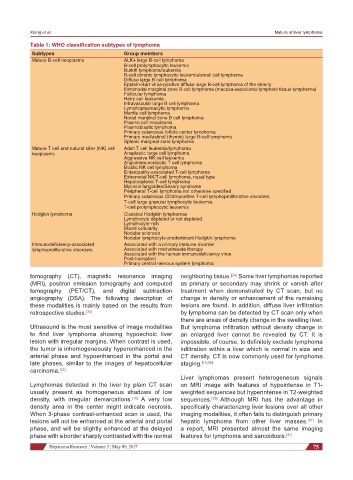
Table 1: WHO classification subtypes of lymphoma
Subtypes Group members
Mature B-cell neoplasms ALK+ large B-cell lymphoma
B-cell prolymphocytic leukemia
Burkitt lymphoma/leukemia
B-cell chronic lymphocytic leukemia/small cell lymphoma
Diffuse large B cell lymphoma
Epstein-Barr virus-positive diffuse large B-cell lymphoma of the elderly
Extranodal marginal zone B cell lymphoma (mucosa-associated lymphoid tissue lymphoma)
Follicular lymphoma
Hairy cell leukemia
Intravascular large B cell lymphoma
Lymphoplasmacytic lymphoma
Mantle cell lymphoma
Nodal marginal zone B cell lymphoma
Plasma cell neoplasms
Plasmablastic lymphoma
Primary cutaneous follicle center lymphoma
Primary mediastinal (thymic) large B-cell lymphoma
Splenic marginal zone lymphoma
Mature T cell and natural killer (NK) cell Adult T cell leukemia/lymphoma
neoplasms Anaplastic large cell lymphoma
Aggressive NK cell leukemia
Angioimmunoblastic T cell lymphoma
Blastic NK cell lymphoma
Enteropathy-associated T-cell lymphoma
Extranodal NK/T-cell lymphoma, nasal type
Hepatosplenic T-cell lymphoma
Mycosis fungoides/Sezary syndrome
Peripheral T-cell lymphoma not otherwise specified
Primary cutaneous CD30-positive T-cell lymphoproliferative disorders
T-cell large granular lymphocytic leukemia
T-cell prolymphocytic leukemia
Hodgkin lymphoma Classical Hodgkin lymphomas
Lymphocyte depleted or not depleted
Lymphocyte-rich
Mixed cellularity
Nodular sclerosis
Nodular lymphocyte-predominant Hodgkin lymphoma
Immunodeficiency-associated Associated with a primary immune disorder
lymphoproliferative disorders Associated with methotrexate therapy
Associated with the human immunodeficiency virus
Post-transplant
Primary central nervous system lymphoma
tomography (CT), magnetic resonance imaging neighboring tissue. [24] Some liver lymphomas reported
(MRI), positron emission tomography and computed as primary or secondary may shrink or vanish after
tomography (PET/CT), and digital subtraction treatment when demonstrated by CT scan, but no
angiography (DSA). The following description of change in density or enhancement of the remaining
these modalities is mainly based on the results from lesions are found. In addition, diffuse liver infiltration
retrospective studies. [34] by lymphoma can be detected by CT scan only when
there are areas of density change in the swelling liver.
Ultrasound is the most sensitive of image modalities But lymphoma infiltration without density change in
to find liver lymphoma showing hypoechoic liver an enlarged liver cannot be revealed by CT. It is
lesion with irregular margins. When contrast is used, impossible, of course, to definitely exclude lymphoma
the tumor is inhomogeneously hyperenhanced in the infiltration within a liver which is normal in size and
arterial phase and hypoenhanced in the portal and CT density. CT is now commonly used for lymphoma
late phases, similar to the images of hepatocellular staging. [15,36]
carcinoma. [35]
Liver lymphomas present heterogeneous signals
Lymphomas detected in the liver by plain CT scan on MRI image with features of hypointense in T1-
usually present as homogeneous shadows of low weighted sequences but hyperintense in T2-weighted
density, with irregular demarcations. [16] A very low sequences. [33] Although MRI has the advantage in
density area in the center might indicate necrosis. specifically characterizing liver lesions over all other
When 3-phase contrast-enhanced scan is used, the imaging modalities, it often fails to distinguish primary
lesions will not be enhanced at the arterial and portal hepatic lymphoma from other liver masses. [37] In
phase, and will be slightly enhanced at the delayed a report, MRI presented almost the same imaging
phase with a border sharply contrasted with the normal features for lymphoma and sarcoidosis. [11]
Hepatoma Research ¦ Volume 3 ¦ May 09, 2017 75