
Page 243 - Read Online
P. 243
A B
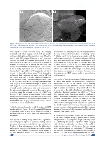
Figure 4: A 64-year-old man with hepatocellular carcinoma in the S2 of the liver. (A) Conventional digital subtraction angiography didn’t show
tumor stain, affected by heart pulsation; (B) coronal multiple planar reconstruction image of cone-beam computed tomography showed a
hypervascular tumor with a diameter of 14 mm in the liver (arrow)
With regard to nodule detection ability, DSA imaging three-dimensional imaging (MIP and VR images) facilitates
combined with CBCT imaging detected all 39 hepatic the easy isolation of anteroposterior overlapping vessels.
nodules, including the eight nodules that were difficult to A detailed search for feeding arteries associated with the
[15]
identify with DSA imaging. Miyayama et al. previously nodules may be conducted if tomographic images (MPR) are
reported that small HCC nodules approximately 1 cm in used after understanding the general vessel anatomy with
size, undetected by DSA imaging, were detected with CBCT three-dimensional imaging [Figure 5]. Another advantage
imaging, thus making TACE possible for such cases. The of CBCT imaging is that it helps in freely determining
average nodule diameter in our study was similar to that the most favorable working angle to isolate the feeding
in the study by Miyayama et al. at 13 mm. In contrast, arteries and nodules that are the target of treatment, using
[15]
Kakeda et al. reported that CBCT imaging did not detect three-dimensional images as a reference. This also makes
[16]
tumors but detected feeding arteries. This is believed to three-dimensional CBCT images useful in superselective
be because their evaluation was based only on MIP and catheterization.
VR images, with small or weakly enhanced nodules not
being revealed during three-dimensional reconstruction. The number of feeding arteries identified by CBCT imaging
In this study, evaluation was made using three-dimensional in caudate lobe nodules has also increased. Endovascular
MPR images as well as MIP and VR images. Evaluation with intervention for HCC in the caudate lobe is generally
[20]
MPR images is considered necessary for the identification considered technically difficult. Caudate branches are
of small nodules and nodules with weak enhancement. high in number and variation. They branch off from the
The advances in diagnostic imaging technology in recent proximal side of the right or left hepatic arterial trunks, as
years have made fine dynamic imaging with MDCT and well as from the distal branches to the right lobe. [21,22] These
liver-specific MRI contrast agents possible, and the small thin branches arising from thick vessels are difficult to
hepatic nodules identified by these methods are becoming identify with two-dimensional DSA imaging, further making
increasingly subjective to endovascular intervention. It is selective catheterization difficult. In addition, multiple
[17]
believed that CBCT imaging could become a useful tool for caudate branches become feeding arteries. With the
identification of such small nodules. addition of CBCT imaging in such cases, we can appreciate
the detailed anatomical relationship between nodules and
No bias by site was observed in nodule detection with CBCT caudate branches with three-dimensional and tomographic
imaging, although Figure 4 clearly shows that CBCT imaging imaging of the vessel, which is considered useful in selecting
is considered useful in areas where DSA imaging alone is an appropriate working angle.
insufficient because of cardiac pulsations and respiratory
movements. In endovascular intervention for HCC, we place a catheter
in the common hepatic artery (proper or replaced hepatic
With regard to feeding artery identification capabilities, artery) and obtain mapping CBCT images with the purpose
CBCT imaging was superior in identifying feeding arteries of establishing therapeutic objectives. CBCT imaging
associated with nodules located in the right hepatic lobe. with selective catheterization is believed to be useful in
This lobe is deep anteroposteriorly, and the right anterior confirming the territory of the tumor that was stained
and posterior branches of the hepatic artery often overlap, before arterial infusion and embolization, although it is
[21]
leading to poor isolation in two-dimensional images of not routinely performed at our facility because its utility is
the anteroposterior direction. [18,19] The addition of CBCT unclear. In addition, frequent imaging takes time and effort.
234 Hepatoma Research | Volume 2 | August 25, 2016