
Page 155 - Read Online
P. 155
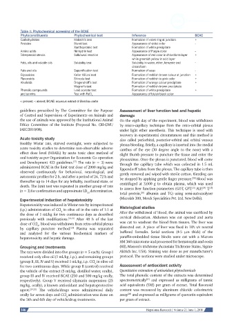
Table 1: Phytochemical screening of the BCAE
           Phytoconstituents           Phytochemical test           Inference                       BCAE
           Carbohydrates               Molisch’s test               Formation of violet ring at junction  +
           Proteins                    Biuret test                  Appearance of violet color      -
                                       Xanthoproteic test           Formation of white precipitate
           Amino acids                 Ninhydrin test               Appearance of Purple color      -
           Triterpenoid sterols        Salkowski reaction           Appearance of red color in chloroform layer   +
                                                                    while greenish yellow in acid layer
           Fats, oils and volatile oils   Solubility test           Solubility in water, ether, benzene and   -
                                                                    chloroform
           Fats and oils               Saponification test          Formation of soap               -
           Glycosides                  Keller Killiani test         Formation of reddish brown colour at junction  +
           Flavonoids                  Shinoda test                 Formation of reddish to pink color  +
           Alkaloids                   Dragendroff’s test           Formation of orange colour precipitate  +
                                       Wagner’s test                Formation of reddish brown precipitate
           Phenolic compounds          Lead acetate test            Formation of white precipitate  +
           and tannins                 Test with FeCl               Appearance of bluish black color
                                                3
           +: present; -: absent; BCAE: aqueous extract of Bombax ceiba
           guidelines  prescribed  by  The  Committee  for  the  Purpose   Assessment of liver function test and hepatic
           of Control and Supervision of Experiments on Animals and   damage
           the use of animals was approved by the Institutional Animal   On the eigth day of the experiment, blood was withdrawn
           Ethics  Committee  of  the  Institute  (Proposal  No.  CRI-GWL/  by micro-capillary technique from the retro-orbital plexus
           IAEC/2010/08).                                     under light ether anesthesia. This technique is used with
                                                              recovery in experimental circumstances and this method is
           Acute toxicity study                               also called periorbital, posterior-orbital and orbital venous
           Healthy  Wistar  rats,  starved  overnight,  were  subjected  to   plexus bleeding. Briefly, a capillary is inserted into the medial
           acute toxicity studies to determine non-observable adverse   canthus of the eye (30 degree angle to the nose) with a
           effect dose level (NOAEL) by acute toxic class method of   slight thumb pressure to puncture the tissue and enter the
           oral toxicity as per Organization for Economic Co-operation   plexus/sinus. Once the plexus is punctured, blood will come
           and Development 423 guidelines.  The rats (n = 3) were   through the capillary tube which was collected in 1.5 mL
                                      [17]
           administered BCAE in the limit test dose of 2000 mg/kg and   Eppendorff tubes from the plexus. The capillary tube is then
           observed continuously for behavioral, neurological, and   gently removed and wiped with sterile cotton. Bleeding can
           autonomic profiles for 2 h, and after a period of 24, 72 h and   be stopped by applying gentle finger pressure.  Blood was
                                                                                                   [20]
           thereafter up to 14 days for any lethality, moribund state, or   centrifuged  at  3,000  g  to  obtain  plasma,  which  was  used
           death. The limit test was repeated in another group of rats   to assess liver function parameters (GOT, GPT,  ALP,  T, [25]
                                                                                                        [24]
                                                                                                   [23]
           (n = 3) for confirmation and approximate LD  determination.
                                                                         [26]
                                             50               total protein,  albumin and TG) using semi-autoanalyser
           Experimental induction of hepatotoxicity           (Microlab 300, Merck Specialities Pvt. Ltd. New Delhi).
           Hepatotoxicity was induced in Wistar rats by intraperitoneal   Histological studies
           (i.p.) administration of CCl  in olive oil in the ratio of 1:1 at
                                4
           the dose of 1 mL/kg for two continuous days as described   After the withdrawal of blood, the animal was sacrificed by
           previously with modifications. [18,19]  After 48 h of the last   cervical dislocation. Abdomen was cut opened and aorta
           dose of CCl , blood was withdrawn from retro-orbital plexus   was cut to washout the blood from tissues. The liver was
                    4
           by capillary puncture method.  Plasma was separated   dissected out. A piece of liver was fixed in 10% v/v neutral
                                     [20]
           and analyzed for the various biochemical markers of   buffered formalin. Serial sections (4-5  μm thick) of the
           hepatotoxicity and hepatic damage.                 paraffin-embedded tissue blocks were cut with a Microm
                                                              HM 360 microtome and processed for hematoxylin and eosin
           Grouping and treatments                            (HE), Masson’s trichrome (Accustain Trichrome Stains, Sigma-
           The rats were divided into five groups (n = 5 each). Group I   Aldrich Inc, USA). Staining was done as per manufacturer’s
           received only olive oil (1 mL/kg, i.p.), and remaining groups   protocol. The sections were studied under microscope.
           (group II, III, IV and V) received 1 mL/kg, i.p. CCl  in olive oil
                                                 4
           for two continuous days. While group II (control) received   Assessment of antioxidant activity
           the vehicle of the extract (5 mL/kg, distilled water, orally),   Quantitative estimation of antioxidant phytochemicals
           group III and IV received BCAE (250 and 500 mg/kg orally,   The total phenolic content of the extracts was determined
                                                                           [27]
           respectively). Group V received silymarin suspension (25   spectrometrically  and expressed as milligrams of tannic
           mg/kg, orally), a known antioxidant and hepatoprotective   acid equivalents (TAE) per gram of extract. Total flavonoid
           agent. [21,22]   The vehicle/drugs were  administered daily   content was measured by aluminum chloride colorimetric
           orally for seven days and CCl administration was done on   assay  and expressed as milligrams of quercetin equivalent
                                                                  [28]
                                   4
           the 5th and 6th day of vehicle/drug treatments.    per gram of extract.
            146                                                            Hepatoma Research | Volume 2 | June 1, 2016