
Page 148 - Read Online
P. 148
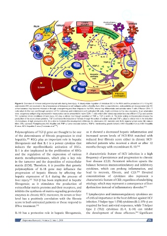
Figure 6: Overview of immune and parenchymal cells during liver injury. A steady-state migration of immature DC to the RLN and the production of IL-10 by KC
            and resident DC are involved in the phenomenon of tolerance to self-antigens within a healthy liver. After a virus infection, viral particles are incorporated into DC
            either because they become infected or through cross-priming and then migrate to the RLN, where they differentiate and activate naive T cells. Effector CD4+ T
            cells return to the liver and through secretion of Th1 cytokines and collaboration with activated NK cells, might contribute to the virus clearance. In an alternative
            view, exogenous antigen (Ag) expressed in hepatocytes can be presented to naive CD8+ T cells which after clonal expansion become efficient CTLs and secrete
            Th1 cytokines Under conditions of liver injury, KC play a critical role through secretion of TNF-α, TGF-β and IL-6. The latter acting on hepatocytes induces the
            production of the acute phase proteins. TGF-β activates the induction of fibrosis through the action of stellate cells and TNF-α plays a critical role in the induction
            of cholestasis. A high production of IL-10 is able to modulate the development of fibrosis. IL: interleukin; DC: dendritic cell; RLN: regional lymph node; NK: natural
            killer; CTL: cytotoxic T lymphocytes; KC: Kupffer cell; TNF-α: tumor necrosis factor-α; TGF-β: transforming growth factor-β; HAV: hepatitis A virus; HBV: hepatitis
            B virus; HCV: hepatitis C virus (Fainboim et al. [104] )
            Polymorphisms of TGF-β gene are thought to be one   as it showed a decreased hepatic inflammation and
            of the determinants of fibrosis progression in viral   increased serum  levels of HCV-RNA  matched with
            hepatitis.  HSCs play an important role in hepatic   reduced liver fibrosis score either  in chronic HCV-
                    [90]
            fibrogenesis and that IL-1 is a potent cytokine that   infected patients who received a short or after 12
            induces  the  myofibroblastic  activation  of  HSCs.   months therapy with recombinant IL-10. [93]
            IL-1 is also implicated in the proliferation of HSCs
            and the regulation of the expression  of various   A characteristic feature of HCV infection is a high
            matrix metalloproteinases, which play a key role   frequency of persistence and progression to chronic
            in the turnover and the deposition of extracellular   liver disease (CLD). Persistent  infection upsets the
            matrix (ECM). Therefore, it is possible that genetic   balance between immunostimulatory and inhibitory
            polymorphism of IL-1B gene may influence the       cytokines,  which can prolong inflammation,  and
                                                                                                   [94]
            progression of hepatic fibrosis by affecting the   lead  to  necrosis,  fibrosis,  and CLD.   Elevated
            hepatic expression  of IL-1  during  the  process  of   concentrations of cytokines also represent a
            liver injury.  TGF-β has been implicated in hepatic   characteristic feature of CLD, regardless of underlying
                      [91]
            fibrogensis; as it stimulates the production  of   etiology, which may represent a consequence of liver
            extracellular matrix proteins and their receptors, and   dysfunction instead of inflammatory disorder. [95]
            inhibits the synthesis of matrix-regrading proteolytic
            enzymes in chronic HCV; moreover, its serum or liver   T lymphocytes and immunoregulatory cytokines are
            level has a positively correlation with the fibrosis   of critical importance in the host defense against HCV
            score in both untreated patients or those respond to   infection. T-helper type 1 (Thl) cytokines (IL-2, IFN-γ) are
            IFN-α treatment. [92]                              required for host anti-viral responses, while T-helper
                                                               type  2 (Th2) cytokines  (IL-4,  IL-10) can inhibit
            IL-10 has a protective  role in  hepatic fibrogenesis,   the  development  of these  effectors. [96]  Significant


                 Hepatoma Research | Volume 2 | June 1, 2016                                              139