
Page 103 - Read Online
P. 103
Post-operative management of normal liver. The deepest ablations were performed
Five patients were transferred to the Intensive Care Unit before the superficial ones to minimize the possibility
and were under observation until patients became stable. of micro bubbles that might obscure visualization of
A naso-gasteric tube was left for 24 h. Patients started the deepest portions of the tumor and thus prevent
oral fluids when intestinal sounds became audible, and complete ablation. In our cases, we ablated the tract
gradually returned to a normal diet. Ambulance was before removal of the needle.
started as early as possible. Drains were removed when
below 100 mL (usually the 4th day). Hospital stay keep Post-ablation care
as short as possible to avoid hospital acquired infection, IV antiemetic was given. Strong IV analgesics were given
ranging from 5 days to 7 days. to control pain. All patients were observed clinically for
2-3 h in the Radiology Department to detect any acute
In the same period, 20 consecutive patients with complications (like bleeding, shock and injury to other
HCC (12 males, 8 females; average age: 54.3 years; organs) and to start IV fluid. Prophylactic antibiotics were
range: 48-66 years) underwent percutaneous RFA at started and continued for 3 days.
Zagazig University Hospitals, Interventional Radiology
Department [Figure 4]. RESULTS
Thirteen of them were treated using the Radionics cool Sociodemographic characteristics of patients
tip needle (4 ablated by the single probe and 8 by the We compared tumor characteristics in the two different
cluster probe). Seven patients were treated using the treatment groups (Child-Pugh score, tumor number,
Rita needle with expandable hooks. Fifteen patients tumor diameter and AFP levels), as shown in Table 1.
were treated with a single electrode insertion, 4 with
double insertions and in one case, by three insertions. Group A: Resection
Only 1 patient received a second session of RFA due A total of 20 consecutive patients with HCC (13 males,
to a residual tumor detected by the 1-month follow-up 7 females; average age: 53.4 years; range: 45-62 years)
triphasic CT study. underwent HR. The etiology of the patients’ underlying
liver disease were characterized by 20 patients with
Local anesthesia was performed on the entry site of the chronic hepatitis (hepatitis B: 3; hepatitis C: 14; hepatitis
skin to the liver capsule along the needle track with 10 mL B + C: 3). On the other hand, 17 had Child A and 3 had
of 2% xylocaine. Most of the patients undergoing RFA were Child B, according to the Child-Pugh scoring system.
treated under general intravenous (IV) anesthesia.
Group B: Radiofrequency ablation
The objective in treating the tumors was to ablate the A total of 20 consecutive patients with HCC (12 males,
entire tumor and an at least 1 cm tumor-free margin 8 females; average age: 54.3 years; range: 48-66
years) underwent RFA interventional in the Radiology
Department. The etiology of the patients’ underlying
liver disease was characterized by 20 patients with
chronic hepatitis (hepatitis B: 4; hepatitis C: 14; hepatitis
B + C: 2). Of these patients, 12 had Child A and 8 Child B.
Table 1: Tumors characteristics in the two different
treatment groups
Underlying cirrhosis Group A Group B
HR (n = 20) (%) RFA (n = 20) (%)
Child-Pugh score
A 17 (85) 12 (60)
B 3 (15) 8 (40)
Number of tumors
Single 18 (90) 13 (65)
Multinodular 2 (10) 7 (35)
Tumor diameter
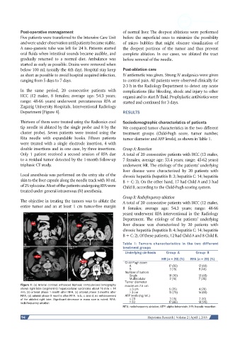
Figure 4: (a) Arterial contrast enhanced triphasic computerized tomography maximum 7.5 cm
shows right lobe (segment 6) hepatocellular carcinoma about 16 mm × 14 ≤ 3 cm 5 (25) 4 (20)
mm; (b) arterial phase 1 month after RFA; (c) arterial phase 3 months after > 3 cm 15 (75) 16 (80)
RFA; (d) arterial phase 9 months after RFA. In b, c and d, no enhancement AFP levels (ng/mL)
of the ablated right lobe. Significant decrease in mass size is noted. RFA: ≤ 20 3 (15) 2 (10)
radiofrequency ablation > 20 17 (85) 18 (90)
RFA: radiofrequency ablation; AFP: alpha-fetoprotein; HR: hepatic resection
94 Hepatoma Research | Volume 2 | April 1, 2016