
Page 9 - Read Online
P. 9
Page 4 of 12 Pedica et al. Hepatoma Res 2021;7:71 https://dx.doi.org/10.20517/2394-5079.2021.89
Figure 4. BilIN (haematoxylin-eosin staining, 20×).
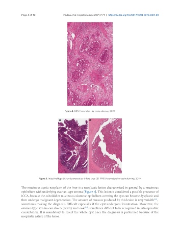
Figure 5. Intestinal type (A) and pancreatico-biliary type (B) IPNB (haematoxylin-eosin staining, 20×).
The mucinous cystic neoplasm of the liver is a neoplastic lesion characterised in general by a mucinous
epithelium with underlying ovarian-type stroma [Figure 7]. This lesion is considered a possible precursor of
iCCA, because the cuboidal or mucinous columnar epithelium covering the cyst can become dysplastic and
[10]
then undergo malignant degeneration. The amount of mucous produced by this lesion is very variable ,
sometimes making the diagnosis difficult especially if the cyst undergoes fenestration. Moreover, the
ovarian-type stroma can also be patchy and loose , sometimes difficult to be recognised in intraoperative
[10]
consultation. It is mandatory to resect the whole cyst once the diagnosis is performed because of the
neoplastic nature of the lesion.