
Page 10 - Read Online
P. 10
Pedica et al. Hepatoma Res 2021;7:71 https://dx.doi.org/10.20517/2394-5079.2021.89 Page 5 of 12
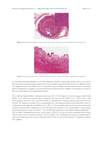
Figure 6. Intraductal tubulopapillary neoplasm of the bile duct (with high-power view) (haematoxylin-eosin staining, 4×).
Figure 7. Mucinous cystic neoplasm of the liver (haematoxylin-eosin staining, 40×) without dysplasia of the epithelium.
In cystoadenocarcinoma [Figure 8], it is often difficult to find the ovarian type stroma, and it is not clear if
they represent a mucinous cystic neoplasm that has undergone regression of the stroma or a different entity.
[1]
The suggested term in the WHO 2019 is mucinous cystic neoplasm with associated invasive carcinoma,
and it is important to recognise it because its behaviour depends on the complete or incomplete resection of
the cyst and the amount of parenchymal invasion.
The combined hepatocellular-cholangiocarcinoma (cHCC-CCA) [Figure 9] is now a category itself inside
[11]
[1]
WHO 2019 with some simplification compared with the previous WHO 2010 . This entity is very
heterogeneous and rare, thus controlled studies to understand its biology and potential treatment are
needed. The diagnosis should rely on morphology first, and immunohistochemistry should be used for
confirmation. Although the real risk factors for cHCC-CCA are largely unknown, they are more commonly
found in cirrhotic liver compared to iCCA, reported to be around 40% in a large series . Some reports
[12]
found that it behaves more as an HCC , but actually their treatment is still without defined guidelines .
[13]
[14]
The so-called “cholangiolocarcinoma” can be a part of cHCC-CCA, but it is more commonly found of part
of iCCA and is now considered a subtype of iCCA [Figure 10] . Differently, the presence of distinct HCC
[1]
and iCCA in the same liver has been reported in fewer than 40 cases in the literature , mainly in cirrhotic
[15]
liver explants.