
Page 8 - Read Online
P. 8
Pedica et al. Hepatoma Res 2021;7:71 https://dx.doi.org/10.20517/2394-5079.2021.89 Page 3 of 12
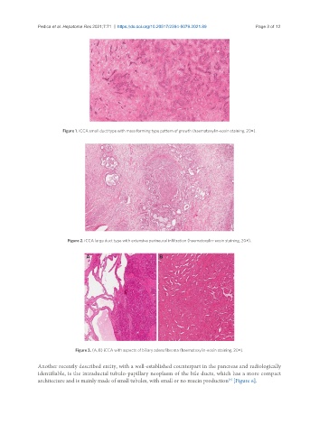
Figure 1. iCCA small duct type with mass forming type pattern of growth (haematoxylin-eosin staining, 20×).
Figure 2. iCCA large duct type with extensive perineural infiltration (haematoxylin-eosin staining, 20×).
Figure 3. (A, B) iCCA with aspects of biliary adenofibroma (haematoxylin-eosin staining, 20×).
Another recently described entity, with a well-established counterpart in the pancreas and radiologically
identifiable, is the intraductal tubulo-papillary neoplasm of the bile ducts, which has a more compact
[9]
architecture and is mainly made of small tubules, with small or no mucin production [Figure 6].