
Page 12 - Read Online
P. 12
Pedica et al. Hepatoma Res 2021;7:71 https://dx.doi.org/10.20517/2394-5079.2021.89 Page 7 of 12
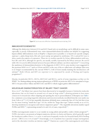
Figure 11. Carcinosarcoma of the liver (haematoxylin-eosin staining, 20×).
IMMUNOHISTOCHEMISTRY
Although the distinction between iCCA and HCC based only on morphology can be difficult in some cases,
especially in poorly differentiated ones, some immunohistochemical markers are helpful for suggesting
hepatocellular differentiation such as HepPar‐1 (hepatocyte in paraffin 1) or hepatocyte specific antigen,
arginase‐1, alpha‐fetoprotein, bile salt export pump and glutamine synthetase. CD10 and pCEA have to
show canalicular pattern of expression. There are no markers specific for biliary differentiation until now,
but CK7 and CK19, although not specific, are usually variably expressed by the biliary tumours. Be careful
[16]
with CK19 in poorly differentiated tumours because different subtype can show expression . Concerning
the usefulness of immunohistochemistry in the diagnosis of cHCC-iCCA, some markers were suggested in
[11]
the previous WHO 2010 such as CD56n and KIT in the case of the so-called stem cell subtype (this term
is no longer recommended by the current WHO ), both of them mainly staining the intermediate cell
[1]
subtype . Both CD56n and KIT are reported to be expressed by canals of Hering and hepatic
[17]
progenitors .
[18]
Mucins, in particular MUC1, MUC2, MUC5AC and MUC6, can be of some importance as they are for
IPMN for distinguishing among immunophenotypes of IPNB, in particular MUC2 for intestinal subtype,
[1]
[7]
MUC1 for pancreatico-biliary and MUC5AC and MUC6 which suggest gastric differentiation .
MOLECULAR CHARACTERIZATION OF BILIARY TRACT CANCER
Since 2014 , the biliary tract cancers have been discovered to be targetable because of distinctive molecular
[19]
alterations that can be found in around 50% of cases in large series , allowing patients to have more
[20]
chances to be treated also when relapsing and opening the way to personalisation of therapies (see Table 1
for a summary concerning targetable alterations and detection methods, modified from the very useful one
from Valle et al. ). iCCAs has targetable alterations definitely more commonly than eCCAs, but this is true
[21]
for the mass forming “small duct type” iCCAs, whilst the “large duct type” behave mostly as an eCCA,
showing mutations in oncogenes and tumour suppressor genes . The targetable alterations mainly regard
[19]
FGFR2 and IDH1 , representing around 20% and 15% of cases, respectively .
[20]
[22]
FGFR2 rearrangements seems to occur mainly in young adults and possibly confer a better prognosis .
[23]
FGFR2 aberrations can present as hotspot mutations, deletion-in-frame and fusions, which generally regard
the breakpoint within intron 17 or exon 18 and more frequently involve the bicaudal family RNA binding
protein 1 (BICC1) in 27.9% of cases . Interestingly, 32.9% of patients have a peculiar unique rearrangement
[24]
and/or the aberration involves an intronic region . Fusions are a part of the rearrangements and induce
[24]