
Page 13 - Read Online
P. 13
Page 8 of 12 Pedica et al. Hepatoma Res 2021;7:71 https://dx.doi.org/10.20517/2394-5079.2021.89
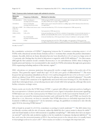
Table 1. Summary table of molecular targets with methods for detection
Molecular Frequency of alteration Methods for detection
target
FGFR alterations 20% of intrahepatic FISH testing for FGFR2 translocation, if available also next-generation DNA sequencing
cholangiocarcinoma cases including FGFR2 intronic region, targeted RNAseq
IDH1/IDH2 15% of intrahepatic DNA sequencing for hotspot mutations in coding region of IDH1 (immunohistochemistry
cholangiocarcinoma cases not applicable)
MSI-high or MMR 5% of biliary tract cancer cases Immunohistochemistry, PCR or tumour next-generation DNA sequencing
deficiency
BRCAness 3.60% Next generation DNA sequencing
ERBB2 (HER2) 1.4%-2.4% Immunohistochemistry and FISH for validation/interpretation of immunohistochemistry to
identify eventual amplification, tumour next-generation DNA sequencing for mutations
BRAF 1% of intrahepatic Immunohistochemistry, DNA sequencing for hotspot mutations in coding region of BRAF
cholangiocarcinoma cases
NTRK 0.75% Immunohistochemistry with Pan-TRK antibodies, next-generation DNA sequencing
including NTRK intronic region or targeted RNAseq, or FISH testing for NTRK translocation
[21]
Modified from Ref .
the constitutive activation of FGFR2 , happening between the N-terminus containing exons 1-19 of
[25]
FGFR2 with a functional tyrosine kinase domain and the C-terminus that contains the partner dimerisation
domain. Recently, deletion-in-frame alteration has also been described in 2.8% of cases in a large series of
[26]
178 patients, also finding that this kind of alteration is targetable with FGFR inhibitors . For this reason,
although the first analysis should consider fluorescence in situ hybridisation (FISH) when looking for
translocation and fusions, it is recommended to also search for FGFR2 aberrations through next-generation
DNA sequencing including analyses of the intronic region .
[21]
IDH1 alterations are missense mutations that generally involve a single residue in the active site of the
enzyme. They generally regard “small duct type” iCCA with poor grade of differentiation [27-29] . The most
frequent hot spot mutations identified are R132C (70%) and less frequently R132L (15%) or R132G (12%) ,
[22]
which are distinct from IDH1 mutant alleles found in glioma and acute myeloid leukaemia . Recently
[30]
Lee et al. found IDH1 mutation to be significantly associated with mass-forming iCCA, bile ductular or
[27]
small duct histological types, not producing mucus and a trend with better prognosis, although this needs
further evaluation on larger series.
Fusion events can involve the NTRK kinase (NTRK 1-3 genes) with different upstream partners, leading to
the overexpression of chimeric protein and constitutively active, ligand-independent downstream signalling.
NTRK fusions are rare in the routine practice for biliary tract cancer. In the recent study by Demols et al. ,
[31]
the cases were screened by immunohistochemistry and confirmed by NGS, and NTRK was found to be
altered in 0.75%. Moreover, we have to remember that NTRK alterations are definitely important for the
[32]
treatment of different malignancies . In the metastatic settings, the guidelines recommend to also directly
apply NGS for detecting NTRK fusions .
[33]
MSI is found in around 5% of iCCAs, sometimes occurring in Lynch syndrome [34,35] . The MSI status does
[35]
not directly correlate with the site of tumour, unless the distal bile duct cancer seems to always be stable
and was found mainly in those with mucinous or papillary morphology . MSI status does have an impact
[35]
[36]
[35]
on survival and is the only predictive marker so far for sensitivity to immune checkpoint inhibitors . In
fact, until now, the immunoexpression of PD-L1 has not been proven to be relevant since it is usually found
in macrophages and not in cancer cells . More studies are needed to investigate this important aim.
[37]