
Page 67 - Read Online
P. 67
Page 8 of 15 Kato et al. Hepatoma Res 2021;7:10 I http://dx.doi.org/10.20517/2394-5079.2020.129
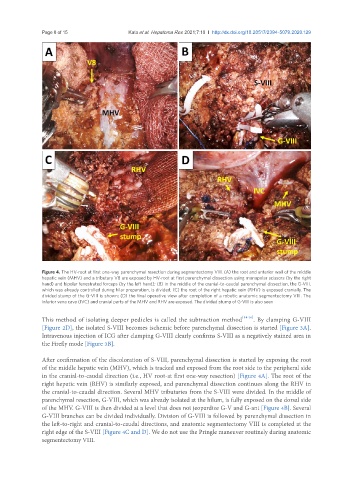
Figure 4. The HV-root at first one-way parenchymal resection during segmentectomy VIII. (A) the root and anterior wall of the middle
hepatic vein (MHV) and a tributary V8 are exposed by HV-root at first parenchymal dissection using monopolar scissors (by the right
hand) and bipolar fenestrated forceps (by the left hand); (B) in the middle of the cranial-to-caudal parenchymal dissection, the G-VIII,
which was already controlled during hilar preparation, is divided; (C) the root of the right hepatic vein (RHV) is exposed cranially. The
divided stump of the G-VIII is shown; (D) the final operative view after completion of a robotic anatomic segmentectomy VIII. The
inferior vena cava (IVC) and cranial parts of the MHV and RHV are exposed. The divided stump of G-VIII is also seen
This method of isolating deeper pedicles is called the subtraction method [14-16] . By clamping G-VIII
[Figure 2D], the isolated S-VIII becomes ischemic before parenchymal dissection is started [Figure 3A].
Intravenous injection of ICG after clamping G-VIII clearly confirms S-VIII as a negatively stained area in
the Firefly mode [Figure 3B].
After confirmation of the discoloration of S-VIII, parenchymal dissection is started by exposing the root
of the middle hepatic vein (MHV), which is tracked and exposed from the root side to the peripheral side
in the cranial-to-caudal direction (i.e., HV root-at first one-way resection) [Figure 4A]. The root of the
right hepatic vein (RHV) is similarly exposed, and parenchymal dissection continues along the RHV in
the cranial-to-caudal direction. Several MHV tributaries from the S-VIII were divided. In the middle of
parenchymal resection, G-VIII, which was already isolated at the hilum, is fully exposed on the dorsal side
of the MHV. G-VIII is then divided at a level that does not jeopardize G-V and G-ant [Figure 4B]. Several
G-VIII branches can be divided individually. Division of G-VIII is followed by parenchymal dissection in
the left-to-right and cranial-to-caudal directions, and anatomic segmentectomy VIII is completed at the
right edge of the S-VIII [Figure 4C and D]. We do not use the Pringle maneuver routinely during anatomic
segmentectomy VIII.