
Page 112 - Read Online
P. 112
Pegoraro et al. Hepatoma Res 2021;7:24 https://dx.doi.org/10.20517/2394-5079.2020.142 Page 3 of 12
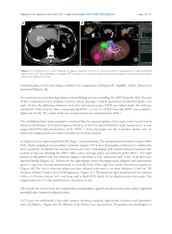
Figure 1. (A) Preoperative CT scan, showing the lesion in segment VIII next to vena cava and the displacement of right and middle
®
hepatic veins. (B) The workstation of Synapse 3D , a software for surgical use that allows the development of virtual reconstructions
based upon triphasic CT scans.
reconstruction of the liver using a software for surgical use (Synapse 3D, Fujifilm, Tokyo, Japan) was
®
®
performed [Figure 1B].
We proposed two resection hypotheses, one including and one excluding the MHV from the FLR. The aim
of this comparison was to evaluate whether venous drainage could be guaranteed by the left hepatic vein
only. In fact, the difference between these two options in terms of FLR was rather small: the software
calculated a FLR of 28.6% when conserving the MHV, vs. a 26.7% of FLR when the MHV was excluded [
Figure 2A and B]. The results of the two reconstructions are summarized in Table 1.
The multidisciplinary team evaluation concluded that the surgical ligation of the right portal branch and its
distal alcoholization, with radiofrequency ablation of the liver parenchyma for right hepatectomy in two
stages (RALPPS) with preservation of the MHV + cholecystectomy was the preferable option, also to
reduce the surgical stress of a major resection in a cirrhotic patient.
In April 2020, the laparoscopic RALPPS stage-1 was performed. The operation started with a biopsy of the
FLR, which confirmed micronodular cirrhosis. Hepatic IOUS were thoroughly performed to confirm the
liver’s anatomy, to identify the known lesions and their relationship with vasculo-biliary structures (the
nodule in Sg8 was abutting the MHV with a clear cleavage plane and infiltrating the RHV). The right
branch of the portal vein was isolated, clipped with Hem-o-Lok, transected and 18 mL of alcohol were
injected distally [Figure 3A]. Afterwards, the right hepatic artery was temporarily clamped, and indocyanine
green (2 mg) was injected intravenously to mark the limit of the right liver under fluorescence guidance
[Figure 3B]. The future resection plane was then ablated with nine 5-cm-deep ablations (Cool-tip™ RF
Ablation System E Series) under IOUS guidance [Figure 3C]. The proximal right portal branch was marked
with a 2/0 Prolene suture, left 4 cm long, and a final IOUS check of the ablation plane was made. The
surgical time was 275 min, and blood loss was about 50 mL.
The patient did not develop any postoperative complication, apart from sinus tachycardia, which regressed
promptly after bisoprolol administration.
A CT scan was performed 2 days after surgery, showing complete right portal occlusion and hypodense
areas of ablation, [Figure 3D]. No dilation of the biliary tract was present. The patient was discharged on