
Page 56 - Read Online
P. 56
Page 6 of 11 Kanmaniraja et al. Hepatoma Res 2020;6:51 I http://dx.doi.org/10.20517/2394-5079.2020.46
A B
C
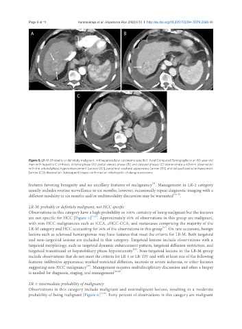
Figure 5. LR-M (Probably or definitely malignant, not hepatocellular carcinoma specific). Axial Computed Tomography in an 80-year-old
man with hepatitis C cirrhosis. Arterial phase (A); portal venous phase (B); and delayed phases (C) demonstrate a 40-mm observation
with rim arterial phase hyperenhancement [arrows (A)], peripheral washout appearance [arrow (B)], and delayed central enhancement
[arrow (C)] observation. Subsequent biopsy confirmed an intrahepatic cholangiocarcinoma
[9]
features favoring benignity and no ancillary features of malignancy . Management in LR-2 category
usually includes routine surveillance in six months; however, occasionally repeat diagnostic imaging with a
different modality in six months and/or multimodality discussion may be warranted [16,18] .
LR-M: probably or definitely malignant, not HCC specific
Observations in this category have a high probability or 100% certainty of being malignant but the features
are not specific for HCC [Figure 5] [7,15] . Approximately 93% of observations in this group are malignant,
with non-HCC malignancies such as iCCA, cHCC-CCA, and metastases comprising the majority of the
LR-M category and HCC accounting for 36% of the observations in this group . On rare occasions, benign
[17]
lesions such as sclerosed hemangiomas may have features that meet the criteria for LR-M. Both targetoid
and non-targetoid lesions are included in this category. Targetoid lesions include observations with a
targetoid morphology, such as targetoid dynamic enhancement pattern, targetoid diffusion restriction, and
targetoid transitional or hepatobiliary phase hypointensity . Non-targetoid lesions in the LR-M group
[19]
include observations that do not meet the criteria for LR-5 or LR-TIV and with at least one of the following
features: infiltrative appearance, marked restricted diffusion, necrosis or severe ischemia, or other features
[19]
suggesting non-HCC malignancy . Management requires multidisciplinary discussion and often a biopsy
is needed for diagnosis, staging, and management [16,18] .
LR-3: intermediate probability of malignancy
Observations in this category include malignant and nonmalignant lesions, resulting in a moderate
probability of being malignant [Figure 6] [7,15] . Forty percent of observations in this category are malignant