
Page 55 - Read Online
P. 55
Kanmaniraja et al. Hepatoma Res 2020;6:51 I http://dx.doi.org/10.20517/2394-5079.2020.46 Page 5 of 11
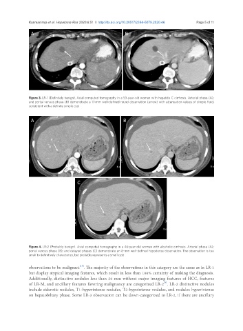
A B
Figure 3. LR-1 (Definitely benign). Axial computed tomography in a 53-year-old woman with hepatitis C cirrhosis. Arterial phase (A);
and portal venous phase (B) demonstrate a 17-mm well-defined round observation (arrow) with attenuation values of simple fluid,
consistent with a definite simple cyst
A B
C
Figure 4. LR-2 (Probably benign). Axial computed tomography in a 46-year-old woman with alcoholic cirrhosis. Arterial phase (A);
portal venous phase (B); and delayed phases (C) demonstrate an 8-mm well-defined hypodense observation. The observation is too
small to definitively characterize, but probably represents a small cyst
[17]
observations to be malignant . The majority of the observations in this category are the same as in LR-1
but display atypical imaging features, which result in less than 100% certainty of making the diagnosis.
Additionally, distinctive nodules less than 20 mm without major imaging features of HCC, features
[9]
of LR-M, and ancillary features favoring malignancy are categorized LR-2 . LR-2 distinctive nodules
include siderotic nodules, T1-hyperintense nodules, T2-hypointense nodules, and nodules hyperintense
on hepatobiliary phase. Some LR-3 observation can be down-categorized to LR-2, if there are ancillary