
Page 58 - Read Online
P. 58
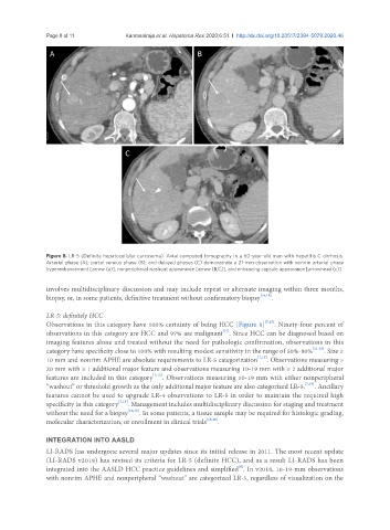
Page 8 of 11                                  Kanmaniraja et al. Hepatoma Res 2020;6:51  I  http://dx.doi.org/10.20517/2394-5079.2020.46

                A                                            B


                                       C


               Figure 8. LR-5 (Definite hepatocellular carcinoma). Axial computed tomography in a 60-year-old man with hepatitis C cirrhosis.
               Arterial phase (A); portal venous phase (B); and delayed phases (C) demonstrate a 21-mm observation with nonrim arterial phase
               hyperenhancement [arrow (a)], nonperipheral washout appearance [arrow (B,C)], and enhancing capsule appearance [arrowhead (c)]

               involves multidisciplinary discussion and may include repeat or alternate imaging within three months,
               biopsy, or, in some patients, definitive treatment without confirmatory biopsy [16,18] .

               LR-5: definitely HCC
               Observations in this category have 100% certainty of being HCC [Figure 8] [7,15] . Ninety-four percent of
                                                                     [17]
               observations in this category are HCC and 97% are malignant . Since HCC can be diagnosed based on
               imaging features alone and treated without the need for pathologic confirmation, observations in this
               category have specificity close to 100% with resulting modest sensitivity in the range of 50%-80% [21-23] . Size ≥
               10 mm and nonrim APHE are absolute requirements to LR-5 categorization [7,15] . Observations measuring >
               20 mm with ≥ 1 additional major feature and observations measuring 10-19 mm with ≥ 2 additional major
               features are included in this category [7,15] . Observations measuring 10-19 mm with either nonperipheral
               “washout” or threshold growth as the only additional major feature are also categorized LR-5. [7,15] . Ancillary
               features cannot be used to upgrade LR-4 observations to LR-5 in order to maintain the required high
               specificity in this category [7,15] . Management includes multidisciplinary discussion for staging and treatment
               without the need for a biopsy [16,18] . In some patients, a tissue sample may be required for histologic grading,
               molecular characterization, or enrollment in clinical trials [16,18] .


               INTEGRATION INTO AASLD
               LI-RADS has undergone several major updates since its initial release in 2011. The most recent update
               (LI-RADS v2018) has revised its criteria for LR-5 (definite HCC), and as a result LI-RADS has been
                                                                          [6]
               integrated into the AASLD HCC practice guidelines and simplified . In v2018, 10-19-mm observations
               with nonrim APHE and nonperipheral “washout” are categorized LR-5, regardless of visualization on the