
Page 57 - Read Online
P. 57
Kanmaniraja et al. Hepatoma Res 2020;6:51 I http://dx.doi.org/10.20517/2394-5079.2020.46 Page 7 of 11
A B
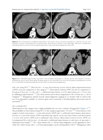
Figure 6. LR-3 (Intermediate probability hepatocellular carcinoma). Axial computed tomography in a 68-year-old man with
hepatitis C cirrhosis. Arterial phase (A); and portal venous phase (B) demonstrate a 12-mm observation with nonrim arterial phase
hyperenhancement [arrow (A)] and no washout appearance or enhancing capsule appearance [arrow (B)]
A B
Figure 7. LR-4 (Probable hepatocellular carcinoma). Axial computed tomography in a 68-year-old man with hepatitis C cirrhosis.
Arterial phase (A); and portal venous phase (B) demonstrate a 9-mm observation with nonrim arterial phase hyperenhancement [arrow
(A)], nonperipheral washout appearance [arrow (B)], and no enhancing capsule appearance
[17]
with 38% being HCC . Observations < 20 mm demonstrating nonrim arterial phase hyperenhancement
(APHE) alone are categorized in this category [7,15] . Observations without APHE can also be categorized in
this group if they are < 20 mm with ≤ 1 additional major feature, or if the observations are ≥ 20 mm with
no additional major features [7,15] . LR-4 observations can be down-categorized to LR-3 if there are ≥ 1 AF of
benignity [7,15] . Management options include repeat diagnostic imaging in 3-6 months with or without the
use of an alternative modality or contrast agent and occasionally a multidisciplinary discussion may be
warranted [16,18] .
LR-4: probably HCC
Observations in this category have a high probability but not 100% certainty of being HCC [Figure 7] [7,15] .
The probability of observations being HCC in this category approaches 75%-80% [17,20] . Observations in this
category include observations < 10 mm with nonrim APHE and ≥ 1 additional major feature, observations
that are 10-19 mm with nonrim APHE and enhancing “capsule” as the only major feature, and observations
≥ 20 mm with nonrim APHE and no additional major feature. Observations without nonrim APHE can
be categorized LR-4 with the size < 20 mm and ≥ 2 additional major features or with size ≥ 20 mm and ≥ 1
additional major feature [7,15] . LR-3 observations with ≥ 1 AF favoring malignancy can be upgraded to LR-4,
[9]
while LR-5 observations with ≥ 1 AF favoring benignity can be downgraded to LR-4 . Management often