
Page 34 - Read Online
P. 34
Garg et al. Hepatoma Res 2019;5:39 I http://dx.doi.org/10.20517/2394-5079.2019.009 Page 3 of 11
n
n n
n n
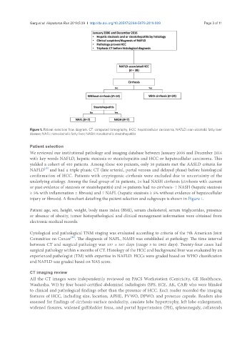
Figure 1. Patient selection flow diagram. CT: computed tomography; HCC: hepatocellular carcinoma; NAFLD: non-alcoholic fatty liver
disease; NAFL: non-alcoholic fatty liver; NASH: nonalcoholic steatohepatitis
Patient selection
We reviewed our institutional pathology and imaging database between January 2006 and December 2016
with key words NAFLD, hepatic steatosis or steatohepatitis and HCC or hepatocellular carcinoma. This
yielded a cohort of 400 patients. Among these 400 patients, only 38 patients met the AASLD criteria for
[15]
NAFLD and had a triple phasic CT (late arterial, portal venous and delayed phase) before histological
confirmation of HCC. Patients with cryptogenic cirrhosis were excluded due to uncertainty of the
underlying etiology. Among the final group of 38 patients, 24 had NASH cirrhosis (cirrhosis with current
or past evidence of steatosis or steatohepatitis) and 14 patients had no cirrhosis- 7 NASH (hepatic steatosis
≥ 5% with inflammation ± fibrosis) and 7 NAFL (hepatic steatosis ≥ 5% without evidence of hepatocellular
injury or fibrosis). A flowchart detailing the patient selection and subgroups is shown in Figure 1.
Patient age, sex, height, weight, body mass index (BMI), serum cholesterol, serum triglycerides, presence
or absence of obesity, tumor histopathological and clinical management information were obtained from
electronic medical records.
Cytological and pathological TNM staging was evaluated according to criteria of the 7th American Joint
[26]
Committee on Cancer . The diagnosis of NAFL, NASH was established at pathology. The time interval
between CT and surgical pathology was 137 ± 387 days (range 3 to 1802 days). Twenty-four cases had
surgical pathology within 6 months of CT. Histology of the HCC and background liver was evaluated by an
experienced pathologist (TM) with expertise in NAFLD. HCCs were graded based on WHO classification
and NAFLD was graded based on NAS score.
CT imaging review
All the CT images were independently reviewed on PACS Workstation (Centricity, GE Healthcare,
Waukesha, WI) by four board-certified abdominal radiologists (SPS, ECE, AK, CAB) who were blinded
to clinical and pathological findings other than the presence of HCC. Each reader recorded the imaging
features of HCC, including size, location, APHE, PVWO, DPWO, and presence capsule. Readers also
assessed for findings of cirrhosis-surface nodularity, caudate lobe hypertrophy, left lobe enlargement,
widened fissures, widened gallbladder fossa, and portal hypertension (PH), splenomegaly, collaterals