
Page 37 - Read Online
P. 37
Page 6 of 11 Garg et al. Hepatoma Res 2019;5:39 I http://dx.doi.org/10.20517/2394-5079.2019.009
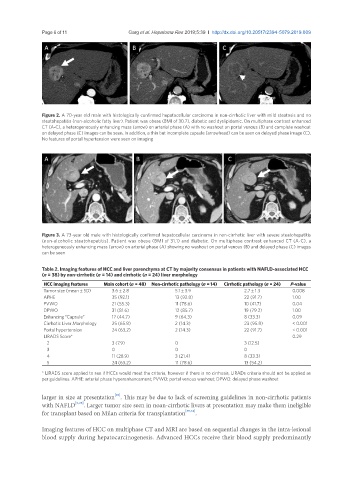
Figure 2. A 70-year old male with histologically confirmed hepatocellular carcinoma in non-cirrhotic liver with mild steatosis and no
steatohepatitis (non-alcoholic fatty liver). Patient was obese (BMI of 30.7), diabetic and dyslipidemic. On multiphase contrast enhanced
CT (A-C), a heterogeneously enhancing mass (arrow) on arterial phase (A) with no washout on portal venous (B) and complete washout
on delayed phase (C) images can be seen. In addition, a thin but incomplete capsule (arrowhead) can be seen on delayed phase image (C).
No features of portal hypertension were seen on imaging
Figure 3. A 73-year old male with histologically confirmed hepatocellular carcinoma in non-cirrhotic liver with severe steatohepatitis
(non-alcoholic steatohepatitis). Patient was obese (BMI of 31.1) and diabetic. On multiphase contrast enhanced CT (A-C), a
heterogeneously enhancing mass (arrow) on arterial phase (A) showing no washout on portal venous (B) and delayed phase (C) images
can be seen
Table 2. Imaging features of HCC and liver parenchyma at CT by majority consensus in patients with NAFLD-associated HCC
(n = 38) by non-cirrhotic (n = 14) and cirrhotic (n = 24) liver morphology
HCC imaging features Main cohort (n = 48) Non-cirrhotic pathology (n = 14) Cirrhotic pathology (n = 24) P-value
Tumor size (mean ± SD) 3.6 ± 2.8 5.1 ± 3.9 2.7 ± 1.3 0.008
APHE 35 (92.1) 13 (92.8) 22 (91.7) 1.00
PVWO 21 (55.3) 11 (78.6) 10 (41.7) 0.04
DPWO 31 (81.6) 12 (85.7) 19 (79.2) 1.00
Enhancing “Capsule” 17 (44.7) 9 (64.3) 8 (33.3) 0.09
Cirrhotic Liver Morphology 25 (65.8) 2 (14.3) 23 (95.8) < 0.001
Portal hypertension 24 (63.2) 2 (14.3) 22 (91.7) < 0.001
LIRADS Score* 0.29
2 3 (7.9) 0 3 (12.5)
3 0 0 0
4 11 (28.9) 3 (21.4) 8 (33.3)
5 24 (63.2) 11 (78.6) 13 (54.2)
* LIRADS score applied to see if HCCs would meet the criteria, however if there is no cirrhosis, LIRADs criteria should not be applied as
per guidelines. APHE: arterial phase hyperenhancement; PVWO: portal venous washout; DPWO: delayed phase washout
[20]
larger in size at presentation . This may be due to lack of screening guidelines in non-cirrhotic patients
with NAFLD [3,28] . Larger tumor size seen in noan-cirrhotic livers at presentation may make them ineligible
for transplant based on Milan criteria for transplantation [35,36] .
Imaging features of HCC on multiphase CT and MRI are based on sequential changes in the intra-lesional
blood supply during hepatocarcinogenesis. Advanced HCCs receive their blood supply predominantly