
Page 38 - Read Online
P. 38
Garg et al. Hepatoma Res 2019;5:39 I http://dx.doi.org/10.20517/2394-5079.2019.009 Page 7 of 11
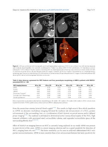
Figure 4. A 47-year old female with histologically confirmed hepatocellular carcinoma (HCC) in non-cirrhotic liver with minimal steatosis
and moderate steatohepatitis (non-alcoholic steatohepatitis). Patient was obese (BMI of 30.5) and diabetic. On multiphase contrast
enhanced CT (A-C), three of the four readers interpreted this as a heterogeneously enhancing mass (arrow) on arterial phase (A) with
no washout on portal venous (B) and delayed phase (C) images. However 1of the four readers interpreted this as a heterogeneously
enhancing mass (arrow) on arterial phase (A) with washout on portal venous (B) and delayed phase (C) images. A resected specimen (D)
showing HCC (arrow) with background fatty liver can be seen
Table 3. Inter-observer agreement for HCC features and liver parenchyma morphology at MRI in patients with NAFLD-
associated HCC (n = 38)
HCC imaging features R1 vs. R2 R1 vs. R3 R1 vs. R4 R2 vs. R3 R2 vs. R4 R3 vs. R4
APHE 0.74 0.79 0.79 0.74 0.84 1.0
PVWO 0.42 0.00 0.53 0.05 0.32 0.05
DPWO 0.95 0.84 0.74 0.79 0.68 0.47
Enhancing “Capsule” 0.47 0.05 0.42 0.37 0.79 0.37
Cirrhotic Liver Morphology 0.79 0.89 0.89 0.79 0.79 0.89
Portal hypertension 0.74 0.79 0.95 0.84 0.79 0.84
Data are presented as prevalence-adjusted bias-adjusted kappa. R1: reader 1; R2: reader 2; R3: reader 3; R4: reader 4; APHE: arterial phase
hyperenhancement; PVWO: portal venous phase washout; DPWO: delayed phase washout
from the anomalous arteries (arterial blood supply) [37,38] . This results in high arterial flow which manifests
as APHE on dynamic multiphase imaging followed by washout (de-enhancement of a HCC, greater
enhancement of the surrounding liver, or a combination of both factors) on portal venous and/or delayed
phase imaging [39-43] . The washout is attributed to diminished portal venous blood supply of the HCC, high
tumoral cellularity with associated small extracellular volume, and expanded extracellular space of the
surrounding fibrotic liver [37,38,43,44] .
Effect of NAFLD on imaging features on HCC is currently being explored. In our study APHE was present
in most (92.1%) of the cases. APHE is a major imaging criterion and has a good sensitivity for detection of
HCC, ranging from 65%-96% [39,43,45] . The lower sensitivity can be seen in early/well differentiated HCC with
partial neovascularization. APHE is more sensitive than other enhancement features but lacks specificity for