


Page 18 - Read Online
P. 18
Page 8 of 12                          Yu et al. Hepatoma Res. 2025;11:29 | https://dx.doi.org/10.20517/2394-5079.2025.47


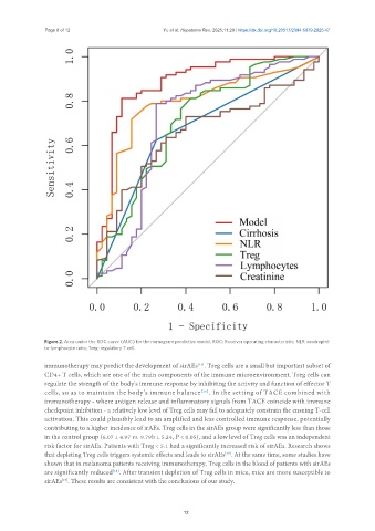
               Figure 2. Area under the ROC curve (AUC) for the nomogram predictive model. ROC: Receiver operating characteristic; NLR: neutrophil-
               to-lymphocyte ratio; Treg: regulatory T cell.

               immunotherapy may predict the development of sirAEs . Treg cells are a small but important subset of
                                                                [15]
               CD4+ T cells, which are one of the main components of the immune microenvironment. Treg cells can
               regulate the strength of the body's immune response by inhibiting the activity and function of effector T
               cells, so as to maintain the body's immune balance [16] . In the setting of TACE combined with
               immunotherapy - where antigen release and inflammatory signals from TACE coincide with immune
               checkpoint inhibition - a relatively low level of Treg cells may fail to adequately constrain the ensuing T-cell
               activation. This could plausibly lead to an amplified and less controlled immune response, potentially
               contributing to a higher incidence of irAEs. Treg cells in the sirAEs group were significantly less than those
               in the control group (6.07 ± 4.97 vs. 9.79b ± 5.24, P < 0.05), and a low level of Treg cells was an independent
               risk factor for sirAEs. Patients with Treg < 5.1 had a significantly increased risk of sirAEs. Research shows
               that depleting Treg cells triggers systemic effects and leads to sirAEs . At the same time, some studies have
                                                                        [17]
               shown that in melanoma patients receiving immunotherapy, Treg cells in the blood of patients with sirAEs
               are significantly reduced . After transient depletion of Treg cells in mice, mice are more susceptible to
                                     [18]
               sirAEs . These results are consistent with the conclusions of our study.
                     [19]

