
Page 234 - Read Online
P. 234
Page 454 Shami-shah et al. Extracell Vesicles Circ Nucleic Acids 2023;4:447-60 https://dx.doi.org/10.20517/evcna.2023.14
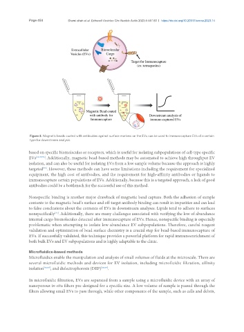
Figure 3. Magnetic beads coated with antibodies against surface markers on the EVs can be used to immunocapture EVs of a certain
type for downstream analysis.
based on specific biomolecules or receptors, which is useful for isolating subpopulations of cell-type specific
EVs [20,47,53] . Additionally, magnetic bead-based methods may be automated to achieve high throughput EV
isolation, and can also be useful for isolating EVs from a low sample volume because the approach is highly
targeted . However, these methods can have some limitations including the requirement for specialized
[53]
equipment, the high cost of antibodies, and the requirement for high-affinity antibodies or ligands to
immunocapture certain populations of EVs. Additionally, because this is a targeted approach, a lack of good
antibodies could be a bottleneck for the successful use of this method.
Nonspecific binding is another major drawback of magnetic bead capture. Both the adhesion of sample
contents to the magnetic bead’s surface and off-target antibody binding can result in impurities and can lead
to false conclusions about the contents of EVs in downstream analyses. Lipids tend to adhere to surfaces
nonspecifically . Additionally, there are many challenges associated with verifying the low of abundance
[27]
internal cargo biomolecules detected after immunocapture of EVs. Hence, nonspecific binding is especially
problematic when attempting to isolate low-abundance EV subpopulations. Therefore, careful reagent
validation and optimization of bead surface chemistry is a crucial step for bead-based immunocapture of
EVs. If successfully validated, this technique provides a powerful platform for rapid immunoenrichment of
both bulk EVs and EV subpopulations and is highly adaptable to the clinic.
Microfluidics-based methods
Microfluidics enable the manipulation and analysis of small volumes of fluids at the microscale. There are
several microfluidic methods and devices for EV isolation, including microfluidic filtration, affinity
isolation [56,57] , and dielectrophoresis (DEP) [58,59] .
In microfluidic filtration, EVs are separated from a sample using a microfluidic device with an array of
nanoporous in-situ filters pre-designed for a specific size. A low volume of sample is passed through the
filters allowing small EVs to pass through, while other components of the sample, such as cells and debris,