
Page 210 - Read Online
P. 210
Page 571 Loh et al. Extracell Vesicles Circ Nucleic Acids 2023;4:568-87 https://dx.doi.org/10.20517/evcna.2023.34
Sorting-at-Entry and involves several mechanistic steps, including aggregation, the use of sorting scaffold/
receptors, and structural motifs (e.g., disulfide bond) [19,20] .
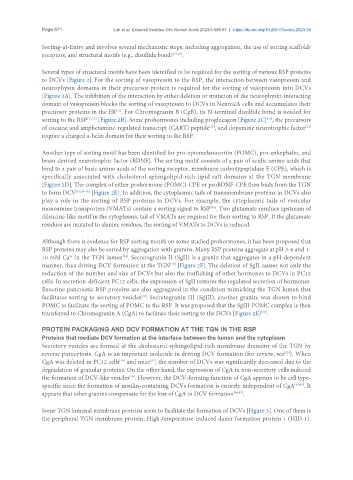
Several types of structural motifs have been identified to be required for the sorting of various RSP proteins
to DCVs [Figure 2]. For the sorting of vasopressin to the RSP, the interaction between vasopressin and
neurophysin domains in their precursor protein is required for the sorting of vasopressin into DCVs
[Figure 2A]. The inhibition of the interaction by either deletion or mutation of the neurophysin-interacting
domain of vasopressin blocks the sorting of vasopressin to DCVs in Neuro2A cells and accumulates their
[21]
precursor proteins in the ER . For Chromogranin B (CgB), its N-terminal disulfide bond is needed for
sorting to the RSP [22,23] [Figure 2B]. Some prohormones including proglucagon [Figure 2C] , the precursors
[24]
[25]
[26]
of cocaine and amphetamine-regulated transcript (CART) peptide , and dopamine neurotrophic factor
require a charged α-helix domain for their sorting to the RSP.
Another type of sorting motif has been identified for pro-opiomelanocortin (POMC), pro-enkephalin, and
brain-derived neurotrophic factor (BDNF). The sorting motif consists of a pair of acidic amino acids that
bind to a pair of basic amino acids of the sorting receptor, membrane carboxypeptidase E (CPE), which is
specifically associated with cholesterol-sphingolipid-rich lipid raft domains at the TGN membrane
[Figure 2D]. The complex of either prohormone (POMC)-CPE or proBDNF-CPE then buds from the TGN
to form DCV [17,28-34] [Figure 2E]. In addition, the cytoplasmic tails of transmembrane proteins in DCVs also
play a role in the sorting of RSP proteins to DCVs. For example, the cytoplasmic tails of vesicular
monoamine transporters (VMATs) contain a sorting signal to RSP . Two glutamate residues upstream of
[35]
dileucine-like motif in the cytoplasmic tail of VMATs are required for their sorting to RSP. If the glutamate
residues are mutated to alanine residues, the sorting of VMATs to DCVs is reduced.
Although there is evidence for RSP sorting motifs on some studied prohormones, it has been proposed that
RSP proteins may also be sorted by aggregation with granins. Many RSP proteins aggregate at pH 5-6 and 1-
2+
[36]
10 mM Ca in the TGN lumen . Secretogranin II (SgII) is a granin that aggregates in a pH-dependent
manner, thus driving DCV formation at the TGN [Figure 2F]. The deletion of SgII causes not only the
[37]
reduction of the number and size of DCVs but also the trafficking of other hormones to DCVs in PC12
cells. In secretion-deficient PC12 cells, the expression of SgII restores the regulated secretion of hormones.
Exocrine pancreatic RSP proteins are also aggregated in the condition mimicking the TGN lumen that
[38]
facilitates sorting to secretory vesicles . Secretogranin III (SgIII), another granin, was shown to bind
POMC to facilitate the sorting of POMC to the RSP. It was proposed that the SgIII-POMC complex is then
[39]
transferred to Chromogranin A (CgA) to facilitate their sorting to the DCVs [Figure 2E] .
PROTEIN PACKAGING AND DCV FORMATION AT THE TGN IN THE RSP
Proteins that mediate DCV formation at the interface between the lumen and the cytoplasm
Secretory vesicles are formed at the cholesterol-sphingolipid-rich membrane domains of the TGN by
reverse pinocytosis. CgA is an important molecule in driving DCV formation (for review, see ). When
[15]
CgA was deleted in PC12 cells and mice , the number of DCVs was significantly decreased due to the
[41]
[40]
degradation of granular proteins. On the other hand, the expression of CgA in non-secretory cells induced
the formation of DCV-like vesicles . However, the DCV-forming function of CgA appears to be cell type-
[40]
specific since the formation of insulin-containing DCVs formation is entirely independent of CgA [42,43] . It
appears that other granins compensate for the loss of CgA in DCV formation [44,45] .
Some TGN lumenal membrane proteins seem to facilitate the formation of DCVs [Figure 3]. One of them is
the peripheral TGN membrane protein, High-temperature-induced dauer formation protein 1 (HID-1).