


Page 97 - Read Online
P. 97
Siddiqui et al. Chem Synth 2023;3:25  https://dx.doi.org/10.20517/cs.2023.02    Page 43 of 50


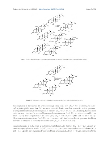
                        Figure 74. Biotransformation of 20-hydroxymethylpregna-1,4-dien-3-one (395) with Cunninghamella elegans.


                              Figure 75. Biotransformation of 6-dehydroprogesterone (395) with Macrophomina phaseolina.

               Hydroxylation in derivatives, 13-hydroxyisolongi-folen-4-one (37) (IC  = 9.64 ± 0.0008 μM) and 9-
                                                                               50
               hydroxyisolongifolen-4-one (40) (IC  = 6.68 ± 0.0096 μM), has increased their activities against tyrosinase,
                                              50
               as compared to substrate, (+)-isolongifolen-4-one (35) (IC  = 51.91 ± 0.0245 μM). Similarly, hydroxylation
                                                                 50
               in derivatives, 17α-ethynyl-11α, 17β-dihydroxyandrost-4-en-3-one (278) (IC  = 5.95 ± 0.00078 μM), 17α-
                                                                                 50
               ethyl-11α, 17β-dihydroxyandrost-4-en-3-one (284) (IC  = 3.46 ± 0.01046 μM), and 17α-ethyl-6α, 17β-
                                                                50
               dihydroxy-5α-androstan-3-one (285) (IC  = 1.72 ± 0.00089 μM) also increased their tyrosinase inhibitory
                                                  50
               activities, as compared to substrate, ethisterone (277) (IC  = 2.61 ± 0.037328 μM).
                                                               50
               Structural changes in metabolites, 4β-methoxycaryophyllene-5α, 14-diol (46) (IC  = 3.09 ± 2.61 μg/mL), 4β-
                                                                                   50
               methoxycaryophyllene-5α, 15-diol (47) (IC  = 0.72 ± 0.17 μg/mL), and caryophyllene-5α,15-diol (48) (IC  =
                                                   50
                                                                                                       50
               1.35 ± 0.43 μg/mL), have significantly increased their anti-malarial activity in vitro, in comparison to the

