
Page 18 - Read Online
P. 18
Page 230 Venkatesh et al. Cancer Drug Resist 2021;4:223-32 I http://dx.doi.org/10.20517/cdr.2020.84
A
B
C
D
E
F
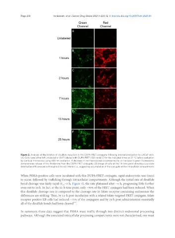
Figure 2. Analysis of the kinetics of disulfide reduction in the DUPA-FRET conjugate following internationalization by LNCaP cells.
(A) Cells were either left unlabeled or (B-F) labeled with DUPA-FRET (100 nmol/L) for the indicated times at 37 °C before evaluation
by confocal microscopy using 488 nm excitation. A decrease in red fluorescence accompanied by an increase in green fluorescence
demonstrates release of the rhodamine from the DUPA-FRET conjugate; (B) image of cells at the 1-h time point showing a punctate
distribution of fluorescence throughout the cell interior; i.e., suggesting accumulation of the conjugate within intracellular compartments
When PSMA-positive cells were incubated with this DUPA-FRET conjugate, rapid endocytosis was found
to occur, followed by trafficking through intracellular compartments. Although the initial rate of disulfide
bond cleavage was fairly rapid (t ~3 h, Figure 3), the rate plateaued after ~6 h, progressing little further
1/2
even out to 24 h. In fact, at the 24-h time point, only ~50% of the FRET conjugate had been reduced. When
this disulfide cleavage rate is compared to the cleavage rate in folate receptor-containing endosomes the
differences are striking. Thus, by 12-h post-incubation with a related folate-targeted FRET conjugate, folate
receptor positive KB cells had reduced ~75% of the conjugates and by 24 h post-administration essentially
[11]
all of the disulfide bonds had been cleaved .
In summary, these data suggest that PSMA must traffic through two distinct endosomal processing
pathways. Although the associated intracellular processing compartments were not characterized, one must