
Page 15 - Read Online
P. 15
Venkatesh et al. Cancer Drug Resist 2021;4:223-32 I http://dx.doi.org/10.20517/cdr.2020.84 Page 227
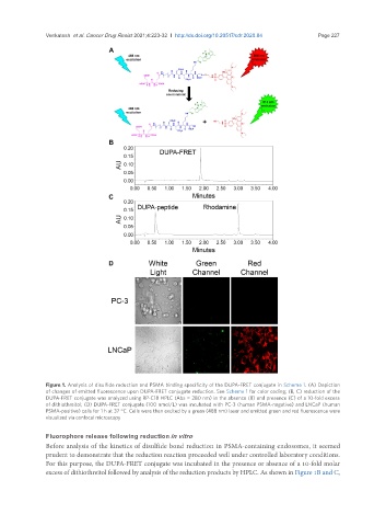
Figure 1. Analysis of disulfide reduction and PSMA binding specificity of the DUPA-FRET conjugate in Scheme 1. (A) Depiction
of changes of emitted fluorescence upon DUPA-FRET conjugate reduction. See Scheme 1 for color coding; (B, C) reduction of the
DUPA-FRET conjugate was analyzed using RP-C18 HPLC (Abs = 280 nm) in the absence (B) and presence (C) of a 10-fold excess
of dithiothreitol; (D) DUPA-FRET conjugate (100 nmol/L) was incubated with PC-3 (human PSMA-negative) and LNCaP (human
PSMA-positive) cells for 1 h at 37 °C. Cells were then excited by a green (488 nm) laser and emitted green and red fluorescence were
visualized via confocal microscopy
Fluorophore release following reduction in vitro
Before analysis of the kinetics of disulfide bond reduction in PSMA-containing endosomes, it seemed
prudent to demonstrate that the reduction reaction proceeded well under controlled laboratory conditions.
For this purpose, the DUPA-FRET conjugate was incubated in the presence or absence of a 10-fold molar
excess of dithiothreitol followed by analysis of the reduction products by HPLC. As shown in Figure 1B and C,