
Page 88 - Read Online
P. 88
Page 14 of 20 Choi et al. Cancer Drug Resist. 2026;9:12
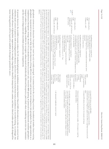
Anesthetized (ketamine + dexdomitor) • Increased NP delivery across BTB/BBB
› IV inj. of FL-labeled NPs (Cy5; 1 μg/g) & MBs
• Mice (male, athymic, › US treatment (5 d p.t.i.) • Whole-brain: 3.5×/4.5× higher FL signal at 0.45/0.55 MPa
nude) › Ex vivo FL imaging (6 h p.t.; euthanized, brain harvested, tumor [MB] • Dissected tumor: 2.3× higher FL signal at 0.55 MPa; no significant increase at 0.45
• Glioma (IC, U87) • Albumin-shelled MPa
dissected)
5
› Ex vivo confocal micro. • 1 × 10 per gram body • Increased NP delivery into tumor microvessels and tissue
weight
Anesthetized (ketamine + dexdomitor) [Transducer]
› IV inj. of luciferase-bearing NPs (1 μg/g) & MBs • Focused • Enhanced transgene expression in both tumor models: ~4× increases in total flux
› US treatment (5 d/7 d p.t.i. for glioma/melanoma) • Fc: 1.1 MHz and average radiance
• Mice (male, athymic, › Ex vivo BL imaging (3 d p.t.; IP inj. of luciferin, euthanized at 5 min [Sonication]
nude) p.i., tumor harvested) • PNP: 0.45/0.55 MPa in
Curley et water
al. [65] • Glioma (IC, U87-mCherry) Anesthetized (ketamine + dexdomitor) • Duty cycle: 0.5% (10 ms
• Melanoma (IC, › IV inj. of MRI contrast agent (50 μL) & MBs • Pre-US MRI: tumor visible via contrast-agent leakage from tumor vessels
B16-F1-OVA) › Pre-US MRI pulses, 2 s interval) • Post-US MRI: increased contrast enhancement, indicating BBB/BTB disruption
• Eight sonication spots
› US treatment (5 d/7 d p.t.i. for glioma/melanoma) • Flow changes: ~2× increase in velocity magnitude; marked change in direction;
› Re-inj. of MRI contrast agent increased convection transport
› Post-US MRI (0, 4, 8, 12 min p.t.; 3T, T1w)
Anesthetized (ketamine + dexdomitor) Only changes:
› IV inj. of MBs [Transducer]
• Mice (male, athymic, › US treatment (16 d p.t.i.) • Fc: 1 MHz
nude) › CED inj. of ZsGreen-NPs (19 μg/20 μL; 0.33 μL/min) [Sonication] • ~2× increased transfection volume in tumor
• Glioma (IC, U87-mCherry) 81
› Ex vivo confocal micro. (2 d p.i.; euthanized, perfused, brain • PNP: 0.45 MPa
harvested, frozen-sectioned) • Duration: 2 min
US: Ultrasound; ICM: intracisterna magna; inj.: injection; MRI: magnetic resonance imaging; h: hour; p.i.: post-injection; T1w: T1-weighted; FL: fluorescence; micro.: microscopy; MB: microbubble; Fc: central frequency; FWHM:
full-width at half-maximum; MI: mechanical index; PNP: peak negative pressure; min: minute; PVS: perivascular space; AD: Alzheimer disease; ALS: amyotrophic lateral sclerosis; IV: intravenous; p.t.: post-treatment; FLAIR:
fluid-attenuated inversion recovery; BBB: blood-brain barrier; SAS: subarachnoid space; IN: intranasal; inst.: instillation; p.inst.: post-instillation; αSMA: alpha smooth muscle actin; GFAP: glial fibrillary acidic protein; P: pressure;
PRF: pulse repetition frequency; IS: interstitial space; TRPV4: transient receptor potential vanilloid-4; AQP4: aquaporin-4; CaM: calmodulin; dcLNs: deep cervical lymph nodes; I SPTA : spatial-peak temporal-average intensity; CSF:
cerebrospinal fluid; GFP: green fluorescence protein; IC: intracranial; NPs: nanoparticles; d: day; p.t.i.: post tumor implantation; BL: bioluminescence; IP: intraperitoneal; BTB: blood-tumor barrier; CED: convection-enhanced
delivery.
glymphatic system as an alternative therapeutic route has only recently emerged. As summarized in the preceding sections, foundational studies characterizing
glymphatic impairment in tumors may help clarify mechanisms of drug delivery resistance and motivate investigations into glymphatic flow augmentation as a
complementary therapeutic strategy. Because US has already been shown to achieve safe, reversible BBB opening, its potential to simultaneously modulate glymphatic
transport makes it a compelling candidate for synergistic enhancement of intrathecally delivered therapeutics, particularly when considering molecular size constraints
and the extent of dispersion into interstitial or perivascular compartments.
At present, however, the available human evidence for US-mediated glymphatic modulation remains limited. Clinical MRI observations to date are derived from
relatively small cohorts of patients with neurodegenerative diseases, and direct evidence in human brain tumor populations is currently lacking. Accordingly, the
translational relevance of glymphatic augmentation for oncologic drug delivery should be interpreted with caution, and future clinical studies specifically designed for
tumor-bearing patients will be required to establish its therapeutic potential in cancer.